The ALKBH1 Monoclonal Antibody (CAB9221) is a high-quality antibody developed for reliable detection and analysis of target proteins. This antibody, produced in rabbits, exhibits high specificity and sensitivity for detecting ALKBH1 in human samples and has been validated for use in Western blot applications. By binding specifically to the ALKBH1 protein, this antibody enables precise detection and analysis in various cell types, making it an essential tool for studies in molecular biology and cancer research.ALKBH1 is a crucial enzyme involved in repairing alkylated DNA and RNA damage, which is essential for maintaining genomic integrity and preventing mutations that can lead to cancer and other diseases.
This antibody is validated for use in WB, IHC-P, IP, ELISA applications and has demonstrated reactivity against Human, Mouse, Rat samples.
Product Name:
ALKBH1 Monoclonal Antibody
SKU:
CAB9221
Size:
20μL, 100μL
Reactivity:
Human, Mouse, Rat
Clone Number:
ARC2511
Conjugate:
Unconjugated
Immunogen:
Synthetic peptide. This information is considered to be commercially sensitive.
0.5μg-4μg antibody for 200μg-400μg extracts of whole cells
IHC-P
1:200 - 1:2000
ELISA
Recommended starting concentration is 1 μg/mL. Please optimize the concentration based on your specific assay requirements.
Synonyms:
ABH, ABH1, alkB, hABH, ALKBH, ALKBH1
Positive Sample:
293T, A-549, K-562, Neuro-2a, PC-12
Cellular Localization:
Mitochondrion, Nucleus.
Calculated MW:
44kDa
Observed MW:
44kDa
This gene encodes a homolog to the E. coli alkB gene product. The E. coli alkB protein is part of the adaptive response mechanism of DNA alkylation damage repair. It is involved in damage reversal by oxidative demethylation of 1-methyladenine and 3-methylcytosine.
Purification Method
Affinity purification
Gene ID
8846
RRID
AB_2863689
Buffer Information
Store at -20℃. Avoid freeze / thaw cycles. Buffer: PBS containing 50% glycerol and 0.05% BSA, preserved with proclin300 or sodium azide, pH 7.3.
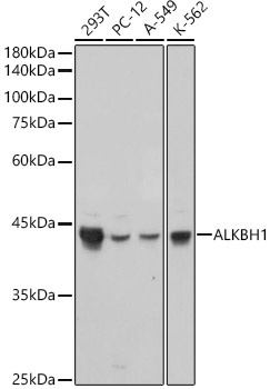
Western blot analysis of various lysates, using ALKBH1 Rabbit mAb (CAB9221) at 1:1000 dilution. Secondary antibody: HRP-conjugated Goat anti-Rabbit IgG (H+L) (CABS014) at 1:10000 dilution. Lysates/proteins: 25μg per lane. Blocking buffer: 3% nonfat dry milk in TBST. Detection: ECL Basic Kit (AbGn00020). Exposure time: 1s.
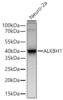
Western blot analysis of lysates from Neuro-2a cells using ALKBH1 Rabbit mAb (CAB9221) at 1:1000 dilution incubated overnight at 4℃. Secondary antibody: HRP-conjugated Goat anti-Rabbit IgG (H+L) (CABS014) at 1:10000 dilution. Lysates/proteins: 25 μg per lane. Blocking buffer: 3% nonfat dry milk in TBST. Detection: ECL Basic Kit (AbGn00020). Exposure time: 45s.
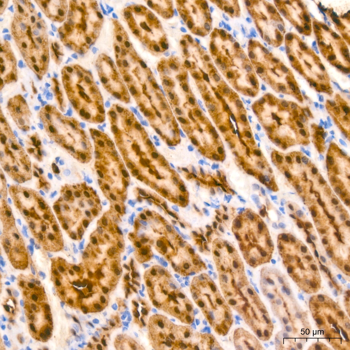
Immunohistochemistry analysis of paraffin-embedded Rat kidney tissue using ALKBH1 Rabbit mAb (CAB9221) at a dilution of 1:200 (40x lens). High pressure antigen retrieval performed with 0.01M Citrate buffer (pH 6.0) prior to IHC staining.
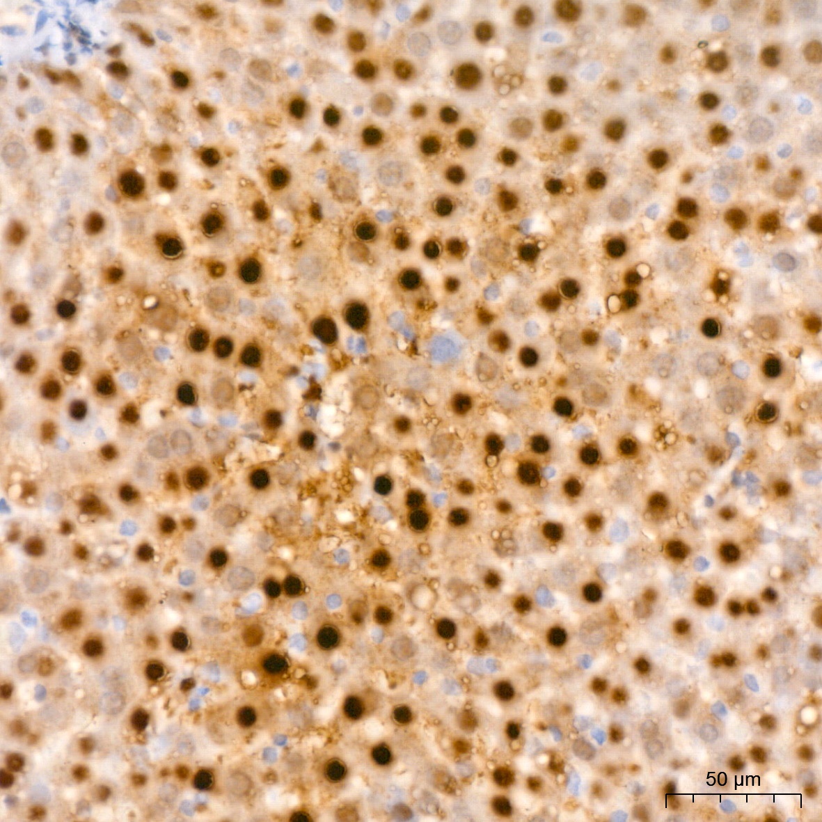
Immunohistochemistry analysis of paraffin-embedded Rat liver tissue using ALKBH1 Rabbit mAb (CAB9221) at a dilution of 1:200 (40x lens). High pressure antigen retrieval performed with 0.01M Citrate buffer (pH 6.0) prior to IHC staining.


![Anti-ALKBH1 [R04-9X6] Monoclonal Antibody (AGMB03274)](https://cdn11.bigcommerce.com/s-h68l9z2lnx/images/stencil/590x590/products/274563/677275/anti-alkbh1-r04-9x6-monoclonal-antibody-agmb03274__46934.1773032080.jpg?c=2 "Anti-ALKBH1 [R04-9X6] Monoclonal Antibody (AGMB03274)")
![Anti-ALKBH1 [R04-4C5] Monoclonal Antibody (AGMB00814)](https://cdn11.bigcommerce.com/s-h68l9z2lnx/images/stencil/590x590/products/272103/691643/anti-alkbh1-r04-4c5-monoclonal-antibody-agmb00814__50683.1774503673.jpg?c=2 "Anti-ALKBH1 [R04-4C5] Monoclonal Antibody (AGMB00814)")


