The ACC1 Monoclonal Antibody (CAB19627) is a high-quality antibody developed for reliable detection and analysis of target proteins. This antibody, raised in rabbits, is highly specific to human samples and has been validated for use in Western blot applications. It binds to the Acc1 protein, allowing for detection and analysis in various cell types, making it an ideal tool for studies in metabolism and lipid biology.Acc1, also known as Acetyl CoA Carboxylase 1, plays a crucial role in fatty acid synthesis and regulation of lipid metabolism.
This antibody is validated for use in WB, IHC-P, IP, ELISA applications and has demonstrated reactivity against Human, Mouse, Rat samples.
Product Name:
ACC1 Monoclonal Antibody
SKU:
CAB19627
Size:
20μL, 100μL
Reactivity:
Human, Mouse, Rat
Clone Number:
ARC2201
Conjugate:
Unconjugated
Immunogen:
Synthetic peptide. This information is considered to be commercially sensitive.
Actin Cytoskeleton, Cytosol, Fibrillar Center, Mitochondrion.
Calculated MW:
266kDa
Observed MW:
277kDa
Acetyl-CoA carboxylase (ACC) is a complex multifunctional enzyme system. ACC is a biotin-containing enzyme which catalyzes the carboxylation of acetyl-CoA to malonyl-CoA, the rate-limiting step in fatty acid synthesis. There are two ACC forms, alpha and beta, encoded by two different genes. ACC-alpha is highly enriched in lipogenic tissues. The enzyme is under long term control at the transcriptional and translational levels and under short term regulation by the phosphorylation/dephosphorylation of targeted serine residues and by allosteric transformation by citrate or palmitoyl-CoA. Multiple alternatively spliced transcript variants divergent in the 5' sequence and encoding distinct isoforms have been found for this gene.
Purification Method
Affinity purification
Gene ID
31
Buffer Information
Store at -20℃. Avoid freeze / thaw cycles. Buffer: PBS containing 50% glycerol and 0.05% BSA, preserved with proclin300 or sodium azide, pH 7.3.
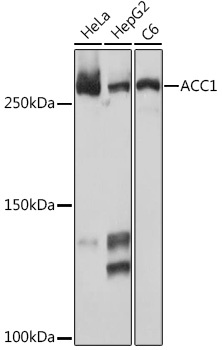
Western blot analysis of various lysates using ACC1 Rabbit mAb (CAB19627) at 1:1000 dilution. Secondary antibody: HRP-conjugated Goat anti-Rabbit IgG (H+L) (CABS014) at 1:10000 dilution. Lysates/proteins: 25μg per lane. Blocking buffer: 3% nonfat dry milk in TBST. Detection: ECL Basic Kit (AbGn00020). Exposure time: 10s.
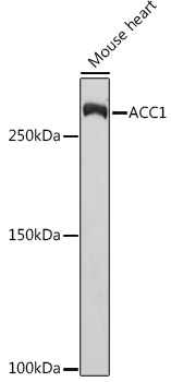
Western blot analysis of lysates from Mouse heart, using ACC1 Rabbit mAb (CAB19627) at 1:1000 dilution. Secondary antibody: HRP-conjugated Goat anti-Rabbit IgG (H+L) (CABS014) at 1:10000 dilution. Lysates/proteins: 25μg per lane. Blocking buffer: 3% nonfat dry milk in TBST. Detection: ECL Basic Kit (AbGn00020). Exposure time: 30s.
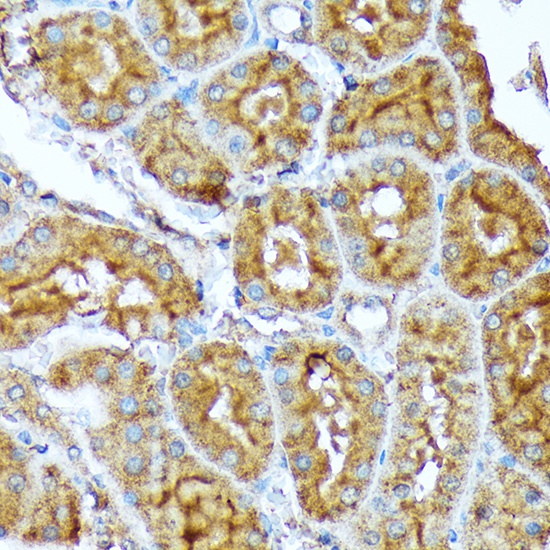
Immunohistochemistry analysis of paraffin-embedded Rat kidney using ACC1 Rabbit mAb (CAB19627) at dilution of 1:100 (40x lens). Microwave antigen retrieval performed with 0.01M Tris/EDTA Buffer (pH 9.0) prior to IHC staining.
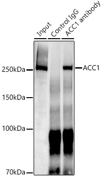
Immunoprecipitation analysis of 300 μg extracts of HepG2 cells using 3 μg ACC1 antibody (CAB19627). Western blot was performed from the immunoprecipitate using ACC1 (CAB19627) at a dilution of 1:1000.


![Anti-Phospho-ACC1 (Ser79) [R05-3D6] Monoclonal Antibody (AGMB05070)](https://cdn11.bigcommerce.com/s-h68l9z2lnx/images/stencil/590x590/products/276355/676433/anti-phospho-acc1-ser79-r05-3d6-monoclonal-antibody-agmb05070__68338.1773029423.jpg?c=2 "Anti-Phospho-ACC1 (Ser79) [R05-3D6] Monoclonal Antibody (AGMB05070)")


