The CD35/CR1 Monoclonal Antibody (CAB3661) is a high-quality antibody developed for reliable detection and analysis of target proteins. This antibody, produced and validated in rabbits, exhibits high specificity and sensitivity towards human samples, making it suitable for various research applications, including Western blotting and immunohistochemistry.CD35, also known as Complement Receptor 1 (CR1), plays a crucial role in the immune system by recognizing and clearing pathogens and immune complexes. Dysregulation of CD35/CR1 has been implicated in various autoimmune disorders, infections, and inflammatory conditions.
This antibody is validated for use in IHC-P, ELISA, IF-P, mIHC applications and has demonstrated reactivity against Human samples.
Product Name:
CD35/CR1 Monoclonal Antibody
SKU:
CAB3661
Size:
20μL, 100μL
Reactivity:
Human
Clone Number:
ARC2065
Conjugate:
Unconjugated
Immunogen:
Synthetic peptide. This information is considered to be commercially sensitive.
Recommended starting concentration is 1 μg/mL. Please optimize the concentration based on your specific assay requirements.
Synonyms:
KN, C3BR, C4BR, CD35, CD35/CR1
Cellular Localization:
Membrane, Single-Pass Type I Membrane Protein.
Calculated MW:
224kDa
This gene is a member of the receptors of complement activation (RCA) family and is located in the 'cluster RCA' region of chromosome 1. The genome is polymorphic at this locus with allele-specific splice variants encoding different isoforms, based on the presence/absence of long homologous repeats (LHRs). The gene encodes a monomeric single-pass type I membrane glycoprotein found on erythrocytes, leukocytes, glomerular podocytes, and splenic follicular dendritic cells. The Knops blood group system is a system of antigens located on this protein. The protein mediates cellular binding to particles and immune complexes that have activated complement. Decreases in expression of this protein and/or mutations in this gene have been associated with gallbladder carcinomas, mesangiocapillary glomerulonephritis, systemic lupus erythematosus, sarcoidosis and Alzheimer's disease. Mutations in this gene have also been associated with a reduction in Plasmodium falciparum rosetting, conferring protection against severe malaria.
Purification Method
Affinity purification
Gene ID
1378
RRID
AB_2863107
Buffer Information
Store at -20℃. Avoid freeze / thaw cycles. Buffer: PBS containing 50% glycerol and 0.05% BSA, preserved with proclin300 or sodium azide, pH 7.3.
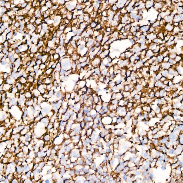
Immunohistochemistry analysis of paraffin-embedded Human tonsil using CD35/CR1 Rabbit mAb (CAB3661) at dilution of 1:50 (40x lens). High pressure antigen retrieval performed with 0.01M Citrate buffer (pH 6.0) prior to IHC staining.
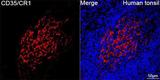
Confocal imaging of paraffin-embedded Human tonsil tissue using CD35/CR1 Rabbit mAb (CAB3661, dilution 1:100) followed by a further incubation with Cy3 Goat Anti-Rabbit IgG (H+L) (CABS007, dilution 1:500) (Red). DAPI was used for nuclear staining (Blue). Objective: 40x. Perform high pressure antigen retrieval with 0.01 M citrate buffer (pH 6.0) prior to IF staining.
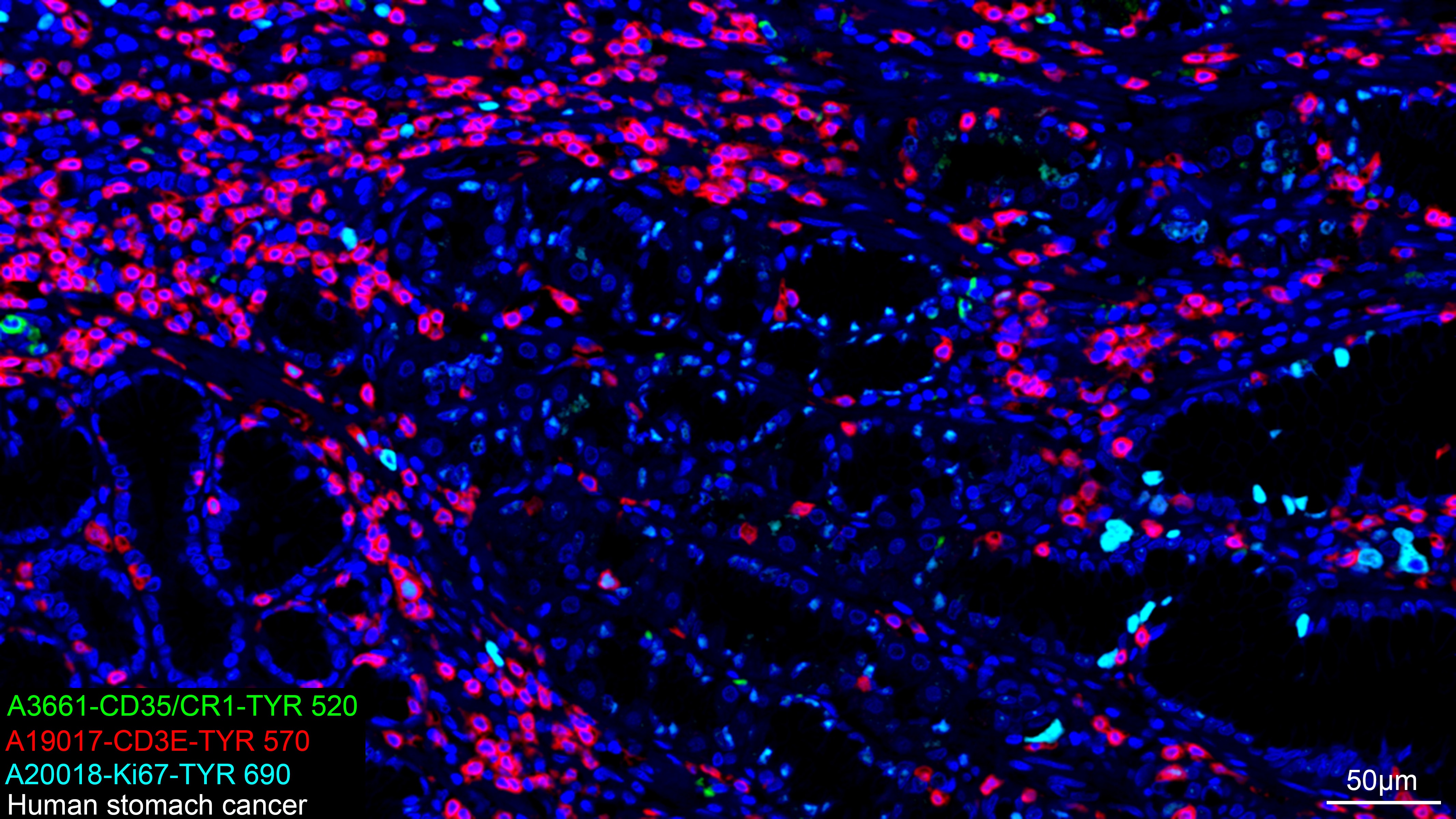
The multiplex IHC analysis on paraffin-embedded Human stomach cancer tissue using the following specific primary antibodies and tyramide signal amplification (TSA) reagents (RK05903) : CD35/CR1 Rabbit mAb (CAB3661, 1:100) with TSA-TYR-520 (Green), CD3E Rabbit mAb (CAB19017, 1:2000) with TSA-TYR-570 (Red), and Ki67 Rabbit mAb (CAB20018, 1:500) with TSA-TYR-690 (cyan). DAPI (Blue) was used for nuclear staining. Prior to multiplex IHC staining, high-pressure antigen retrieval was performed using 0.01M citrate buffer at pH 6.0. The analysis was completed using a 20x objective lens.


![Anti-CD35 [R06-6D1] Monoclonal Antibody (AGMB02040)](https://cdn11.bigcommerce.com/s-h68l9z2lnx/images/stencil/590x590/products/273329/678260/anti-cd35-r06-6d1-monoclonal-antibody-agmb02040__61763.1773035189.jpg?c=2 "Anti-CD35 [R06-6D1] Monoclonal Antibody (AGMB02040)")
![Anti-CD35 [R06-2N9] Monoclonal Antibody (AGMB02995)](https://cdn11.bigcommerce.com/s-h68l9z2lnx/images/stencil/590x590/products/274284/677249/anti-cd35-r06-2n9-monoclonal-antibody-agmb02995__63297.1773032059.jpg?c=2 "Anti-CD35 [R06-2N9] Monoclonal Antibody (AGMB02995)")
![Anti-CD35 [R06-4A-3] Monoclonal Antibody (AGMB06526)](https://cdn11.bigcommerce.com/s-h68l9z2lnx/images/stencil/590x590/products/277807/733565/anti-cd35-r06-4a-3-monoclonal-antibody-agmb06526__90970.1777190375.jpg?c=2 "Anti-CD35 [R06-4A-3] Monoclonal Antibody (AGMB06526)")
![Anti-CD35 [6H10-8B7-9G10] Monoclonal Antibody (AGMB06059)](https://cdn11.bigcommerce.com/s-h68l9z2lnx/images/stencil/590x590/products/277340/677528/anti-cd35-6h10-8b7-9g10-monoclonal-antibody-agmb06059__28370.1773032901.jpg?c=2 "Anti-CD35 [6H10-8B7-9G10] Monoclonal Antibody (AGMB06059)")

