The Cytokeratin 2e Monoclonal Antibody (CAB2615) is a high-quality antibody developed for reliable detection and analysis of target proteins. This antibody, produced in rabbits, is highly specific to human samples and has been validated for use in various applications, including immunohistochemistry and Western blotting.Cytokeratin 2E is a marker for epithelial differentiation and is commonly used in studies related to skin diseases, cancer, and tissue regeneration. The Anti-Cytokeratin 2E Antibody binds specifically to this protein, allowing for accurate detection and analysis in a range of cell types and tissue samples.
This antibody is validated for use in WB, IHC-P, ELISA, IF-P applications and has demonstrated reactivity against Human, Mouse, Rat samples.
Product Name:
Cytokeratin 2e Monoclonal Antibody
SKU:
CAB2615
Size:
20μL, 100μL
Reactivity:
Human, Mouse, Rat
Clone Number:
ARC1925
Conjugate:
Unconjugated
Immunogen:
Recombinant protein (or fragment).This information is considered to be commercially sensitive.
The protein encoded by this gene is a member of the keratin gene family. The type II cytokeratins consist of basic or neutral proteins which are arranged in pairs of heterotypic keratin chains coexpressed during differentiation of simple and stratified epithelial tissues. This type II cytokeratin is expressed largely in the upper spinous layer of epidermal keratinocytes and mutations in this gene have been associated with bullous congenital ichthyosiform erythroderma. The type II cytokeratins are clustered in a region of chromosome 12q12-q13.
Purification Method
Affinity purification
Gene ID
3849
Buffer Information
Store at -20℃. Avoid freeze / thaw cycles. Buffer: PBS containing 50% glycerol and 0.05% BSA, preserved with proclin300 or sodium azide, pH 7.3.
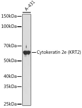
Western blot analysis of lysates from A-431 cells, using Cytokeratin 2e (KRT2) Rabbit mAb (CAB2615) at 1:1000 dilution. Secondary antibody: HRP-conjugated Goat anti-Rabbit IgG (H+L) (CABS014) at 1:10000 dilution. Lysates/proteins: 25μg per lane. Blocking buffer: 3% nonfat dry milk in TBST. Detection: ECL Basic Kit (AbGn00020). Exposure time: 180s.
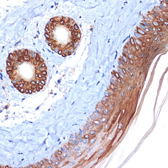
Immunohistochemistry analysis of paraffin-embedded Mouse skin using Cytokeratin 2e (KRT2) Rabbit mAb (CAB2615) at dilution of 1:100 (40x lens). Microwave antigen retrieval performed with 0.01M Tris/EDTA Buffer (pH 9.0) prior to IHC staining.
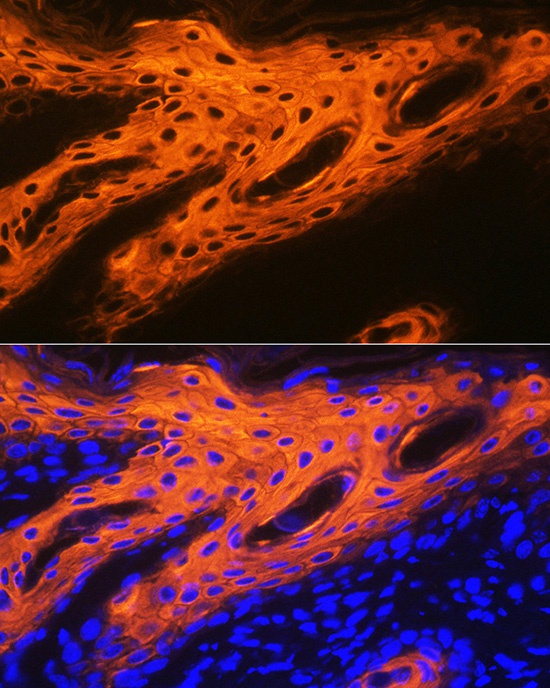
Immunofluorescence analysis of paraffin-embedded human skin using Cytokeratin 2e (KRT2) Rabbit mAb (CAB2615) at dilution of 1:100 (40x lens). Secondary antibody: Cy3-conjugated Goat anti-Rabbit IgG (H+L) (CABS007) at 1:500 dilution. Blue: DAPI for nuclear staining.
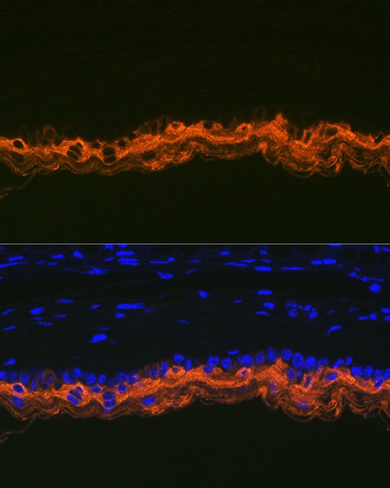
Immunofluorescence analysis of paraffin-embedded mouse skin using Cytokeratin 2e (KRT2) Rabbit mAb (CAB2615) at dilution of 1:100 (40x lens). Secondary antibody: Cy3-conjugated Goat anti-Rabbit IgG (H+L) (CABS007) at 1:500 dilution. Blue: DAPI for nuclear staining.


![Anti-Cytokeratin 2e [R08-2H5] Monoclonal Antibody (AGMB02727)](https://cdn11.bigcommerce.com/s-h68l9z2lnx/images/stencil/590x590/products/274016/680944/anti-cytokeratin-2e-r08-2h5-monoclonal-antibody-agmb02727__70957.1773043697.jpg?c=2 "Anti-Cytokeratin 2e [R08-2H5] Monoclonal Antibody (AGMB02727)")

