The DNPH1 Monoclonal Antibody (CAB2382) is a high-quality antibody developed for reliable detection and analysis of target proteins. This polyclonal antibody, produced in rabbits, exhibits strong reactivity with human samples and has been validated for use in various applications, including Western blotting. By targeting the DNPH1 protein, this antibody enables precise detection and analysis in different cell types, making it an invaluable tool for investigations in genetics, genomics, and cancer biology.
This antibody is validated for use in WB, IF/ICC, ELISA applications and has demonstrated reactivity against Human, Mouse samples.
Product Name:
DNPH1 Monoclonal Antibody
SKU:
CAB2382
Size:
20μL, 100μL
Reactivity:
Human, Mouse
Clone Number:
ARC2571
Conjugate:
Unconjugated
Immunogen:
Synthetic peptide. This information is considered to be commercially sensitive.
Recommended starting concentration is 1 μg/mL. Please optimize the concentration based on your specific assay requirements.
Synonyms:
RCL, C6orf108, dJ330M21.3, DNPH1
Positive Sample:
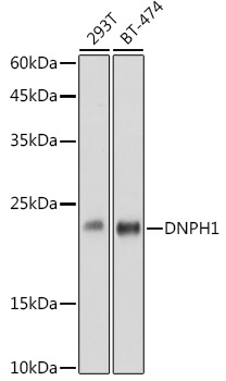
293T, BT-474
Cellular Localization:
Cytosol, Extracellular Exosome, Nucleus.
Calculated MW:
19kDa
Observed MW:
23kDa
This gene was identified on the basis of its stimulation by c-Myc protein. The latter is a transcription factor that participates in the regulation of cell proliferation, differentiation, and apoptosis. The exact function of this gene is not known but studies in rat suggest a role in cellular proliferation and c-Myc-mediated transformation. Two alternative transcripts encoding different proteins have been described.
Purification Method
Affinity purification
Gene ID
10591
Buffer Information
Store at -20℃. Avoid freeze / thaw cycles. Buffer: PBS containing 50% glycerol and 0.05% BSA, preserved with proclin300 or sodium azide, pH 7.3.
Western blot analysis of various lysates using DNPH1 Rabbit mAb (CAB2382) at 1:1000 dilution. Secondary antibody: HRP-conjugated Goat anti-Rabbit IgG (H+L) (CABS014) at 1:10000 dilution. Lysates/proteins: 25μg per lane. Blocking buffer: 3% nonfat dry milk in TBST. Detection: ECL Basic Kit (AbGn00020). Exposure time: 60s.
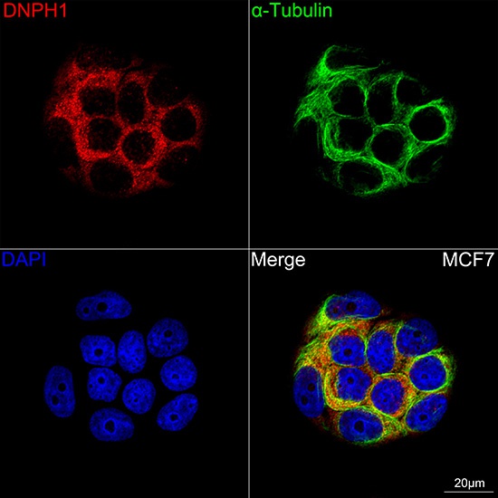
Confocal imaging of MCF7 cells using DNPH1 Rabbit mAb (CAB2382, dilution 1:100) followed by a further incubation with Cy3 Goat Anti-Rabbit IgG (H+L) (CABS007, dilution 1:500) (Red). The cells were counterstained with α-Tubulin Mouse mAb (AC012, dilution 1:400) followed by incubation with ABflo® 488-conjugated Goat Anti-Mouse IgG (H+L) Ab (CABS076, dilution 1:500) (Green). DAPI was used for nuclear staining (Blue). Objective: 100x.