The LEO1 Antibody (CAB8806) is a high-quality antibody developed for reliable detection and analysis of target proteins. This polyclonal antibody, raised in rabbits, is highly specific to human samples and has been validated for use in Western blot and immunohistochemistry applications.LEO1 is essential for proper transcriptional regulation and plays a crucial role in gene expression. Its dysregulation has been linked to various diseases, including cancer and developmental disorders. By targeting LEO1 with this antibody, researchers can investigate its role in transcriptional processes, potentially leading to a better understanding of disease mechanisms and the development of targeted therapies.
This antibody is validated for use in WB, IHC-P, IF/ICC, ELISA applications and has demonstrated reactivity against Human, Mouse, Rat samples.
Product Name:
LEO1 Antibody
SKU:
CAB8806
Size:
20μL, 100μL
Reactivity:
Human, Mouse, Rat
Conjugate:
Unconjugated
Immunogen:
Recombinant protein (or fragment).This information is considered to be commercially sensitive.
Recommended starting concentration is 1 μg/mL. Please optimize the concentration based on your specific assay requirements.
Synonyms:
RDL, LEO1
Positive Sample:
HepG2, Jurkat, Mouse liver, Mouse brain, Rat brain
Cellular Localization:
Nucleus.
Calculated MW:
75kDa
Observed MW:
105kDa
LEO1, parafibromin (CDC73; MIM 607393), CTR9 (MIM 609366), and PAF1 (MIM 610506) form the PAF protein complex that associates with the RNA polymerase II subunit POLR2A (MIM 180660) and with a histone methyltransferase complex (Rozenblatt-Rosen et al., 2005 [PubMed 15632063]).
Purification Method
Affinity purification
Gene ID
123169
RRID
AB_2770166
Buffer Information
Store at -20℃. Avoid freeze / thaw cycles. Buffer: PBS containing 50% glycerol, preserved with proclin300 or sodium azide, pH 7.3.
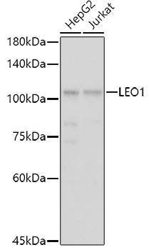
Western blot analysis of various lysates using LEO1 Rabbit pAb (CAB8806) at 1:1000 dilution. Secondary antibody: HRP-conjugated Goat anti-Rabbit IgG (H+L) (CABS014) at 1:10000 dilution. Lysates/proteins: 25μg per lane. Blocking buffer: 3% nonfat dry milk in TBST. Detection: ECL Basic Kit (AbGn00020). Exposure time: 1s.
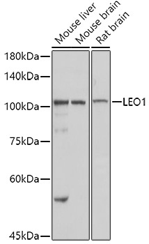
Western blot analysis of various lysates using LEO1 Rabbit pAb (CAB8806) at 1:1000 dilution. Secondary antibody: HRP-conjugated Goat anti-Rabbit IgG (H+L) (CABS014) at 1:10000 dilution. Lysates/proteins: 25μg per lane. Blocking buffer: 3% nonfat dry milk in TBST. Detection: ECL Basic Kit (AbGn00020). Exposure time: 1s.
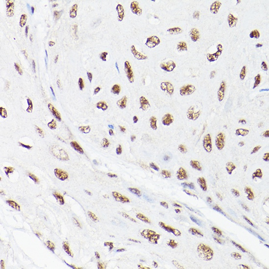
Immunohistochemistry analysis of paraffin-embedded Human esophageal cancer using LEO1 Rabbit pAb (CAB8806) at dilution of 1:100 (40x lens). High pressure antigen retrieval performed with 0.01M Citrate buffer (pH 6.0) prior to IHC staining.
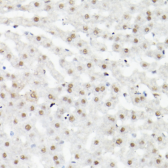
Immunohistochemistry analysis of paraffin-embedded Mouse liver using LEO1 Rabbit pAb (CAB8806) at dilution of 1:100 (40x lens). High pressure antigen retrieval performed with 0.01M Citrate buffer (pH 6.0) prior to IHC staining.
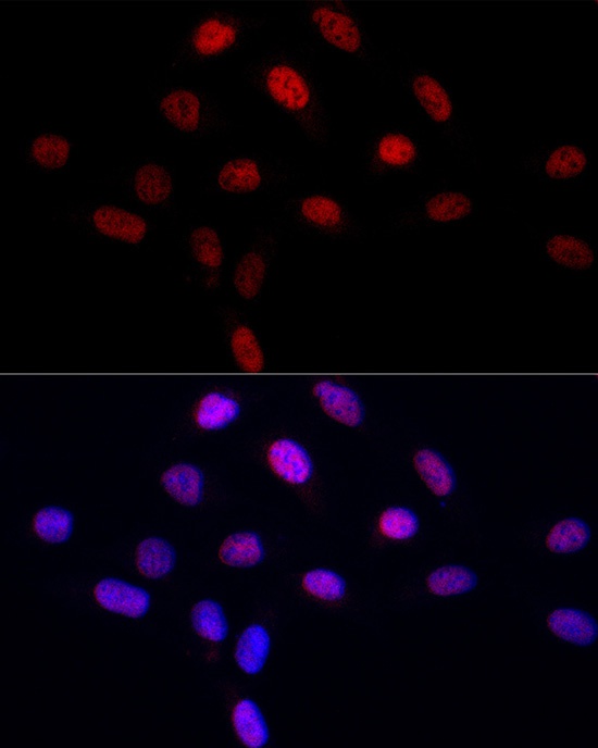
Immunofluorescence analysis of U2OS cells using LEO1 Rabbit pAb (CAB8806) at dilution of 1:50 (40x lens). Secondary antibody: Cy3-conjugated Goat anti-Rabbit IgG (H+L) (CABS007) at 1:500 dilution. Blue: DAPI for nuclear staining.