The LMO2 Monoclonal Antibody (CAB6832) is a high-quality antibody developed for reliable detection and analysis of target proteins. This antibody, raised in rabbits, shows high reactivity with human samples and is validated for use in various applications, including Western blot and immunohistochemistry.LMO2 plays a crucial role in normal hematopoietic development, but its dysregulation is associated with leukemia and other hematological malignancies. Understanding the expression and function of LMO2 in different cell types can provide insights into the mechanisms driving these diseases and potentially lead to the development of targeted therapies.
This antibody is validated for use in WB, ELISA applications and has demonstrated reactivity against Human samples.
Product Name:
LMO2 Monoclonal Antibody
SKU:
CAB6832
Size:
20μL, 100μL
Reactivity:
Human
Clone Number:
ARC1422
Conjugate:
Unconjugated
Immunogen:
Synthetic peptide. This information is considered to be commercially sensitive.
Recommended starting concentration is 1 μg/mL. Please optimize the concentration based on your specific assay requirements.
Synonyms:
TTG2, LMO-2, RBTN2, RHOM2, RBTNL1, LMO2
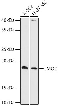
Positive Sample:
K-562, U-87 MG
Cellular Localization:
Nucleoplasm, Nucleus.
Calculated MW:
18kDa
Observed MW:
18kDa
LMO2 encodes a cysteine-rich, two LIM-domain protein that is required for yolk sac erythropoiesis. The LMO2 protein has a central and crucial role in hematopoietic development and is highly conserved. The LMO2 transcription start site is located approximately 25 kb downstream from the 11p13 T-cell translocation cluster (11p13 ttc), where a number T-cell acute lymphoblastic leukemia-specific translocations occur. Alternative splicing results in multiple transcript variants encoding different isoforms.
Purification Method
Affinity purification
Gene ID
4005
Buffer Information
Store at -20℃. Avoid freeze / thaw cycles. Buffer: PBS containing 50% glycerol and 0.05% BSA, preserved with proclin300 or sodium azide, pH 7.3.
Western blot analysis of various lysates using LMO2 Rabbit mAb (CAB6832) at 1:1000 dilution. Secondary antibody: HRP-conjugated Goat anti-Rabbit IgG (H+L) (CABS014) at 1:10000 dilution. Lysates/proteins: 25μg per lane. Blocking buffer: 3% nonfat dry milk in TBST. Detection: ECL Basic Kit (AbGn00020). Exposure time: 90s.


![Anti-LMO2 [R06-5F6] Monoclonal Antibody (AGMB01676)](https://cdn11.bigcommerce.com/s-h68l9z2lnx/images/stencil/590x590/products/272965/676161/anti-lmo2-r06-5f6-monoclonal-antibody-agmb01676__24459.1773028581.jpg?c=2 "Anti-LMO2 [R06-5F6] Monoclonal Antibody (AGMB01676)")
![Anti-LMO2 [R04-4L-7] Monoclonal Antibody (AGMB03661)](https://cdn11.bigcommerce.com/s-h68l9z2lnx/images/stencil/590x590/products/274950/680739/anti-lmo2-r04-4l-7-monoclonal-antibody-agmb03661__92995.1773042996.jpg?c=2 "Anti-LMO2 [R04-4L-7] Monoclonal Antibody (AGMB03661)")


