The MPC1 Antibody (CAB20195) is a high-quality antibody developed for reliable detection and analysis of target proteins. This antibody, raised in rabbits, is highly specific to human samples and is validated for use in Western blot applications. By binding to the MPC1 protein, this antibody allows for the detection and analysis of MPC1 in various cell types, making it an essential reagent for studies in mitochondrial biology and metabolism.MPC1 plays a crucial role in the regulation of glucose metabolism and energy production in the mitochondria.
This antibody is validated for use in WB, IP, ELISA applications and has demonstrated reactivity against Human, Mouse, Rat samples.
Product Name:
MPC1 Antibody
SKU:
CAB20195
Size:
20μL, 100μL
Reactivity:
Human, Mouse, Rat
Conjugate:
Unconjugated
Immunogen:
Synthetic peptide. This information is considered to be commercially sensitive.
0.5μg-4μg antibody for 200μg-400μg extracts of whole cells
ELISA
Recommended starting concentration is 1 μg/mL. Please optimize the concentration based on your specific assay requirements.
Synonyms:
MPYCD, BRP44L, CGI-129, SLC54A1, MPC1
Positive Sample:
HepG2, K-562, Mouse liver, Rat heart
Cellular Localization:
Inner Mitochondrial Membrane Protein Complex, Mitochondrial Inner Membrane, Mitochondrion.
Calculated MW:
12kDa
Observed MW:
12kDa
The protein encoded by this gene is part of an MPC1/MPC2 heterodimer that is responsible for transporting pyruvate into mitochondria. The encoded protein is found in the inner mitochondrial membrane. Defects in this gene are a cause of mitochondrial pyruvate carrier deficiency. Several transcript variants, some protein coding and one non-protein coding, have been found for this gene.
Purification Method
Affinity purification
Gene ID
51660
Buffer Information
Store at -20℃. Avoid freeze / thaw cycles. Buffer: PBS containing 50% glycerol, preserved with proclin300 or sodium azide, pH 7.3.
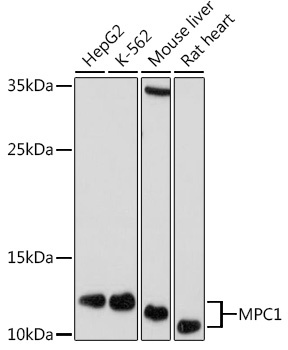
Western blot analysis of various lysates using MPC1 Rabbit pAb (CAB20195) at 1:1000 dilution. Secondary antibody: HRP-conjugated Goat anti-Rabbit IgG (H+L) (CABS014) at 1:10000 dilution. Lysates/proteins: 25μg per lane. Blocking buffer: 3% nonfat dry milk in TBST. Detection: ECL Basic Kit (AbGn00020). Exposure time: 30s.
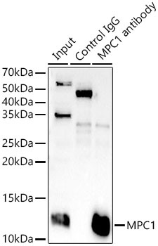
Immunoprecipitation analysis of 300 μg extracts of K-562 cells using 3 μg MPC1 antibody (CAB20195). Western blot was performed from the immunoprecipitate using MPC1 antibody (CAB20195) at a dilution of 1:1000.