The p73 Monoclonal Antibody (CAB2670) is a high-quality antibody developed for reliable detection and analysis of target proteins. This polyclonal antibody, produced in rabbits, is specifically designed for use in Western blot applications and is highly reactive with human samples.The Anti-p73 Antibody binds to the p73 protein, allowing for the detection and analysis of p73 expression in various cell types. This antibody is invaluable for research in fields such as oncology and developmental biology, where the p73 protein is known to be involved in tumor suppression and neuronal development.
This antibody is validated for use in WB, IHC-P, ELISA applications and has demonstrated reactivity against Human, Mouse, Rat samples.
Product Name:
p73 Monoclonal Antibody
SKU:
CAB2670
Size:
20μL, 100μL
Reactivity:
Human, Mouse, Rat
Clone Number:
ARC2628
Conjugate:
Unconjugated
Immunogen:
Synthetic peptide. This information is considered to be commercially sensitive.
Recommended starting concentration is 1 μg/mL. Please optimize the concentration based on your specific assay requirements.
Synonyms:
P73, CILD47, p73
Positive Sample:
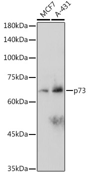
MCF7, A-431
Cellular Localization:
Cytoplasm, Nucleus.
Calculated MW:
70kDa
Observed MW:
80kDa
This gene encodes a member of the p53 family of transcription factors involved in cellular responses to stress and development. It maps to a region on chromosome 1p36 that is frequently deleted in neuroblastoma and other tumors, and thought to contain multiple tumor suppressor genes. The demonstration that this gene is monoallelically expressed (likely from the maternal allele), supports the notion that it is a candidate gene for neuroblastoma. Many transcript variants resulting from alternative splicing and/or use of alternate promoters have been found for this gene, but the biological validity and the full-length nature of some variants have not been determined.
Purification Method
Affinity purification
Gene ID
7161
Buffer Information
Store at -20℃. Avoid freeze / thaw cycles. Buffer: PBS containing 50% glycerol and 0.05% BSA, preserved with proclin300 or sodium azide, pH 7.3.
Western blot analysis of various lysates using p73 Rabbit mAb (CAB2670) at 1:1000 dilution. Secondary antibody: HRP-conjugated Goat anti-Rabbit IgG (H+L) (CABS014) at 1:10000 dilution. Lysates/proteins: 25μg per lane. Blocking buffer: 3% nonfat dry milk in TBST. Detection: ECL Basic Kit (AbGn00020). Exposure time: 180s.
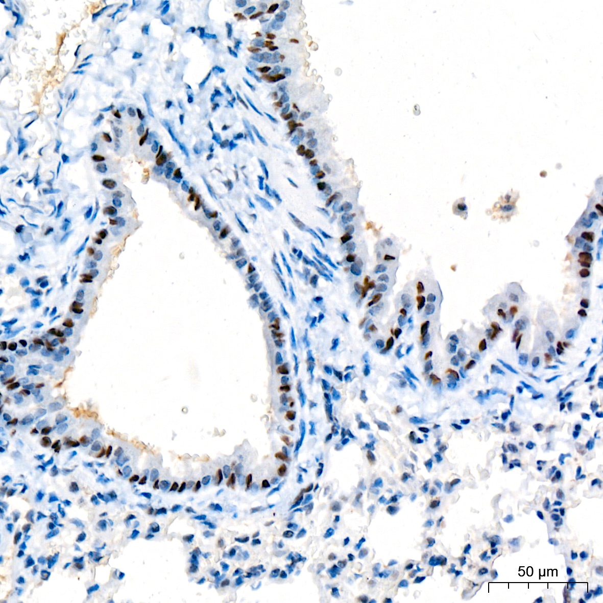
Immunohistochemistry analysis of paraffin-embedded Mouse lung tissue using p73 Rabbit mAb (CAB2670) at a dilution of 1:200 (40x lens). High pressure antigen retrieval was performed with 0.01 M citrate buffer (pH 6.0) prior to IHC staining.
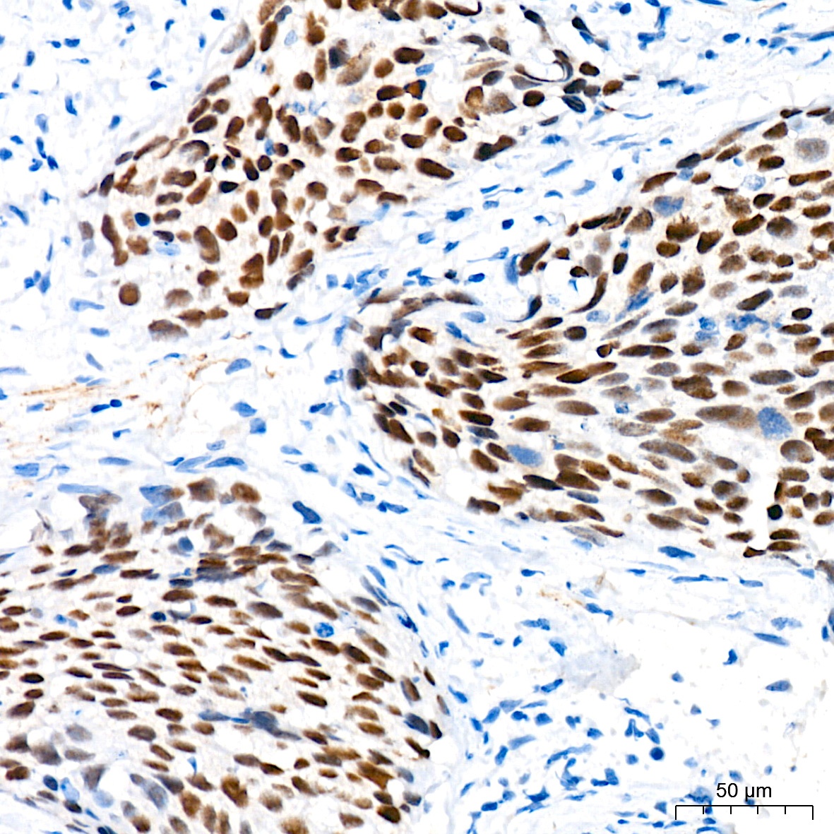
Immunohistochemistry analysis of paraffin-embedded Human cervix cancer tissue using p73 Rabbit mAb (CAB2670) at a dilution of 1:200 (40x lens). High pressure antigen retrieval was performed with 0.01 M citrate buffer (pH 6.0) prior to IHC staining.


![Anti-p73 [R09-1J5] Monoclonal Antibody (AGMB02736)](https://cdn11.bigcommerce.com/s-h68l9z2lnx/images/stencil/590x590/products/274025/679178/anti-p73-r09-1j5-monoclonal-antibody-agmb02736__01170.1773038073.jpg?c=2 "Anti-p73 [R09-1J5] Monoclonal Antibody (AGMB02736)")
![Anti-p73 [R07-1J7] Monoclonal Antibody (AGMB02277)](https://cdn11.bigcommerce.com/s-h68l9z2lnx/images/stencil/590x590/products/273566/678220/anti-p73-r07-1j7-monoclonal-antibody-agmb02277__90135.1773035068.jpg?c=2 "Anti-p73 [R07-1J7] Monoclonal Antibody (AGMB02277)")
![Anti-p73 [R02-3B-2] Monoclonal Antibody (AGMB03703)](https://cdn11.bigcommerce.com/s-h68l9z2lnx/images/stencil/590x590/products/274992/679179/anti-p73-r02-3b-2-monoclonal-antibody-agmb03703__27187.1773038073.jpg?c=2 "Anti-p73 [R02-3B-2] Monoclonal Antibody (AGMB03703)")
![Anti-p73 [R04-6O-3] Monoclonal Antibody (AGMB03893)](https://cdn11.bigcommerce.com/s-h68l9z2lnx/images/stencil/590x590/products/275182/677304/anti-p73-r04-6o-3-monoclonal-antibody-agmb03893__78808.1773032186.jpg?c=2 "Anti-p73 [R04-6O-3] Monoclonal Antibody (AGMB03893)")

