The SERCA1/ATP2A1 Monoclonal Antibody (CAB19639) is a high-quality antibody developed for reliable detection and analysis of target proteins. This antibody, raised in rabbits, has high reactivity with human samples and is validated for use in Western blot applications.SERCA1, also known as ATPase Ca++ transporting cardiac muscle fast-twitch 1, is essential for maintaining calcium homeostasis in muscle cells. Dysregulation of SERCA1 has been linked to various diseases including muscular dystrophy, heart failure, and neurodegenerative disorders.
This antibody is validated for use in WB, ELISA, IF-P applications and has demonstrated reactivity against Mouse, Rat samples.
Product Name:
SERCA1/ATP2A1 Monoclonal Antibody
SKU:
CAB19639
Size:
20μL, 100μL
Reactivity:
Mouse, Rat
Clone Number:
ARC2207
Conjugate:
Unconjugated
Immunogen:
Synthetic peptide. This information is considered to be commercially sensitive.
This gene encodes one of the SERCA Ca(2+)-ATPases, which are intracellular pumps located in the sarcoplasmic or endoplasmic reticula of muscle cells. This enzyme catalyzes the hydrolysis of ATP coupled with the translocation of calcium from the cytosol to the sarcoplasmic reticulum lumen, and is involved in muscular excitation and contraction. Mutations in this gene cause some autosomal recessive forms of Brody disease, characterized by increasing impairment of muscular relaxation during exercise. Alternative splicing results in three transcript variants encoding different isoforms.
Purification Method
Affinity purification
Gene ID
487
Buffer Information
Store at -20℃. Avoid freeze / thaw cycles. Buffer: PBS containing 50% glycerol and 0.05% BSA, preserved with proclin300 or sodium azide, pH 7.3.
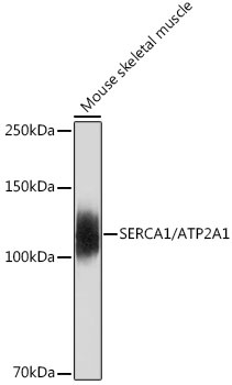
Western blot analysis of lysates from Mouse skeletal muscle, using SERCA1/ATP2A1 Rabbit mAb (CAB19639) at 1:500 dilution. Secondary antibody: HRP-conjugated Goat anti-Rabbit IgG (H+L) (CABS014) at 1:10000 dilution. Lysates/proteins: 25μg per lane. Blocking buffer: 3% nonfat dry milk in TBST. Detection: ECL Basic Kit (AbGn00020). Exposure time: 30s.
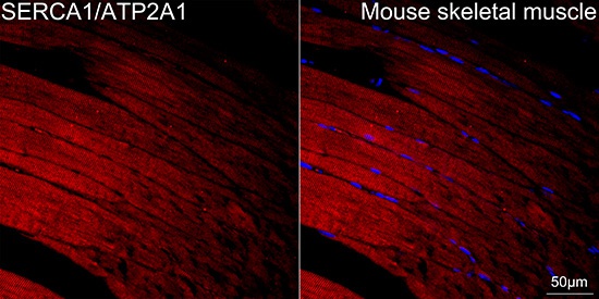
Confocal imaging of paraffin-embedded Mouse skeletal muscle tissue using SERCA1/ATP2A1 Rabbit mAb (CAB19639, dilution 1:100) followed by a further incubation with Cy3 Goat Anti-Rabbit IgG (H+L) (CABS007, dilution 1:500) (Red). DAPI was used for nuclear staining (Blue). High pressure antigen retrieval performed with 0.01M Citrate Buffer (pH 6.0) prior to IF staining. Objective: 40x.


![Anti-SERCA1 ATPase [R07-1X1] Monoclonal Antibody (AGMB03256)](https://cdn11.bigcommerce.com/s-h68l9z2lnx/images/stencil/590x590/products/274545/677799/anti-serca1-atpase-r07-1x1-monoclonal-antibody-agmb03256__02999.1773033752.jpg?c=2 "Anti-SERCA1 ATPase [R07-1X1] Monoclonal Antibody (AGMB03256)")
![Anti-SERCA1 ATPase [R03-2A3] Monoclonal Antibody (AGMB03110)](https://cdn11.bigcommerce.com/s-h68l9z2lnx/images/stencil/590x590/products/274399/676948/anti-serca1-atpase-r03-2a3-monoclonal-antibody-agmb03110__18297.1773031091.jpg?c=2 "Anti-SERCA1 ATPase [R03-2A3] Monoclonal Antibody (AGMB03110)")


