The ARID1B Antibody (CAB15488) is a high-quality antibody developed for reliable detection and analysis of target proteins. This antibody, produced in rabbits, is highly specific to human samples and has been validated for use in Western blotting applications.ARID1B is known to be involved in numerous cellular processes, including cell differentiation, proliferation, and DNA repair. Dysregulation of ARID1B has been implicated in several diseases, including cancer, developmental disorders, and intellectual disabilities.
This antibody is validated for use in WB, IHC-P, IF/ICC, ELISA applications and has demonstrated reactivity against Human, Mouse, Rat samples.
Product Name:
ARID1B Antibody
SKU:
CAB15488
Size:
20μL, 100μL
Reactivity:
Human, Mouse, Rat
Conjugate:
Unconjugated
Immunogen:
Recombinant protein (or fragment).This information is considered to be commercially sensitive.
This locus encodes an AT-rich DNA interacting domain-containing protein. The encoded protein is a component of the SWI/SNF chromatin remodeling complex and may play a role in cell-cycle activation. The protein encoded by this locus is similar to AT-rich interactive domain-containing protein 1A. These two proteins function as alternative, mutually exclusive ARID-subunits of the SWI/SNF complex. The associated complexes play opposing roles. Alternative splicing results in multiple transcript variants.
Purification Method
Affinity purification
Gene ID
57492
RRID
AB_2762887
Buffer Information
Store at -20℃. Avoid freeze / thaw cycles. Buffer: PBS with 0.01% thimerosal,50% glycerol,pH7.3.
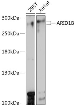
Western blot analysis of various lysates using ARID1B Rabbit pAb (CAB15488) at 1:1000 dilution. Secondary antibody: HRP-conjugated Goat anti-Rabbit IgG (H+L) (CABS014) at 1:10000 dilution. Lysates/proteins: 25μg per lane. Blocking buffer: 3% nonfat dry milk in TBST. Detection: ECL Basic Kit (AbGn00020). Exposure time: 30s.
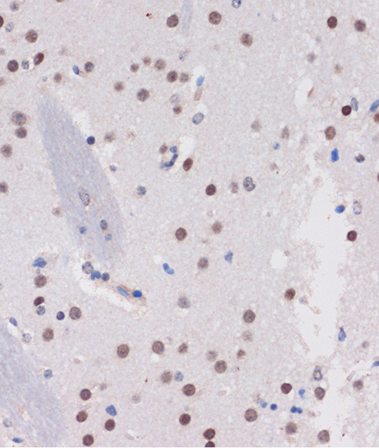
Immunohistochemistry analysis of paraffin-embedded Mouse brain using ARID1B Rabbit pAb (CAB15488) at dilution of 1:100 (40x lens). Microwave antigen retrieval performed with 0.01M PBS Buffer (pH 7.2) prior to IHC staining.
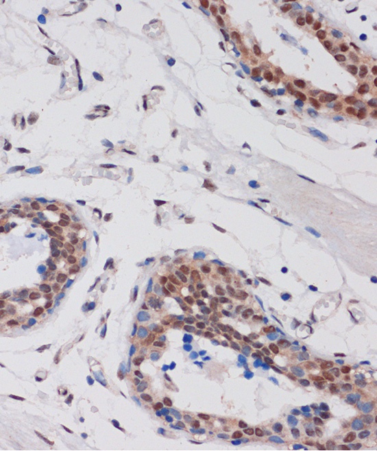
Immunohistochemistry analysis of paraffin-embedded Human breast using ARID1B Rabbit pAb (CAB15488) at dilution of 1:100 (40x lens). Microwave antigen retrieval performed with 0.01M PBS Buffer (pH 7.2) prior to IHC staining.
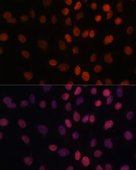
Immunofluorescence analysis of C6 cells using ARID1B Rabbit pAb (CAB15488) at dilution of 1:100. Secondary antibody: Cy3-conjugated Goat anti-Rabbit IgG (H+L) (CABS007) at 1:500 dilution. Blue: DAPI for nuclear staining.