The ATF4 Antibody (CAB0201) is a high-quality antibody developed for reliable detection and analysis of target proteins. This antibody, produced in rabbits, exhibits high reactivity with human samples and has been validated for use in Western blot applications. By binding specifically to ATF4 protein, this antibody allows for accurate detection and analysis in a range of cell types, making it well-suited for investigations in molecular biology and cancer research.ATF4 is known for its role in coordinating cellular responses to stress, such as nutrient deprivation and oxidative stress, by controlling the expression of genes involved in pathways like autophagy and amino acid metabolism.
This antibody is validated for use in WB, IF/ICC, ELISA applications and has demonstrated reactivity against Human samples.
Product Name:
ATF4 Antibody
SKU:
CAB0201
Size:
20μL, 100μL
Reactivity:
Human
Conjugate:
Unconjugated
Immunogen:
Recombinant protein (or fragment).This information is considered to be commercially sensitive.
This gene encodes a transcription factor that was originally identified as a widely expressed mammalian DNA binding protein that could bind a tax-responsive enhancer element in the LTR of HTLV-1. The encoded protein was also isolated and characterized as the cAMP-response element binding protein 2 (CREB-2). The protein encoded by this gene belongs to a family of DNA-binding proteins that includes the AP-1 family of transcription factors, cAMP-response element binding proteins (CREBs) and CREB-like proteins. These transcription factors share a leucine zipper region that is involved in protein-protein interactions, located C-terminal to a stretch of basic amino acids that functions as a DNA binding domain. Two alternative transcripts encoding the same protein have been described. Two pseudogenes are located on the X chromosome at q28 in a region containing a large inverted duplication.
Purification Method
Affinity purification
Gene ID
468
RRID
AB_2757015
Buffer Information
Store at -20℃. Avoid freeze / thaw cycles. Buffer: PBS containing 50% glycerol, preserved with proclin300 or sodium azide, pH 7.3.
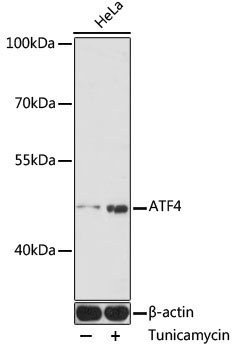
Western blot analysis of lysates from HeLa cells, using ATF4 Rabbit pAb (CAB0201) at 1:1000 dilution. HeLa cells were treated with tunicamycin (2 μg/mL) for 8 hours. Secondary antibody: HRP-conjugated Goat anti-Rabbit IgG (H+L) (CABS014) at 1:10000 dilution. Lysates/proteins: 25μg per lane. Blocking buffer: 3% nonfat dry milk in TBST. Detection: ECL Enhanced Kit (AbGn00021). Exposure time: 3min.
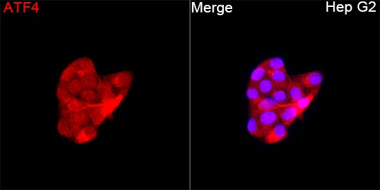
Immunofluorescence analysis of Hep G2 cells using ATF4 Rabbit pAb (CAB0201) at a dilution of 1:100 (40x lens). Secondary antibody: Cy3-conjugated Goat anti-Rabbit IgG (H+L) (CABS007) at 1:500 dilution. Blue: DAPI for nuclear staining.