The ATG13 Antibody (CAB0690) is a high-quality antibody developed for reliable detection and analysis of target proteins. This rabbit-raised antibody is highly specific and reactive with human samples, making it ideal for use in Western blot applications to detect and analyze the ATG13 protein.ATG13 is a key regulator of autophagy, a process that helps cells remove damaged organelles and proteins to maintain cellular health. Dysregulation of autophagy has been implicated in various diseases, including cancer, neurodegenerative disorders, and metabolic diseases.
This antibody is validated for use in WB, IF/ICC, ELISA applications and has demonstrated reactivity against Human, Mouse, Rat samples.
Product Name:
ATG13 Antibody
SKU:
CAB0690
Size:
20μL, 100μL
Reactivity:
Human, Mouse, Rat
Conjugate:
Unconjugated
Immunogen:
Recombinant protein (or fragment).This information is considered to be commercially sensitive.
Recommended starting concentration is 1 μg/mL. Please optimize the concentration based on your specific assay requirements.
Synonyms:
KIAA0652, PARATARG8, ATG13
Positive Sample:
Jurkat, HeLa, U-2 OS, Mouse brain, Mouse testis
Cellular Localization:
Cytoplasm, Preautophagosomal Structure, Cytosol.
Calculated MW:
57kDa
Observed MW:
70kDa
The protein encoded by this gene is an autophagy factor and a target of the TOR kinase signaling pathway. The encoded protein is essential for autophagosome formation and mitophagy.
Purification Method
Affinity purification
Gene ID
9776
RRID
AB_2757341
Buffer Information
Store at -20℃. Avoid freeze / thaw cycles. Buffer: PBS containing 50% glycerol, preserved with proclin300 or sodium azide, pH 7.3.
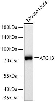
Western blot analysis of lysates from Mouse testis using ATG13 Rabbit pAb (CAB0690) at 1:1000 dilution incubated overnight at 4℃. Secondary antibody: HRP-conjugated Goat anti-Rabbit IgG (H+L) (CABS014) at 1:10000 dilution. Lysates/proteins: 25 μg per lane. Blocking buffer: 3% nonfat dry milk in TBST. Detection: ECL Basic Kit (AbGn00020). Exposure time: 10s.
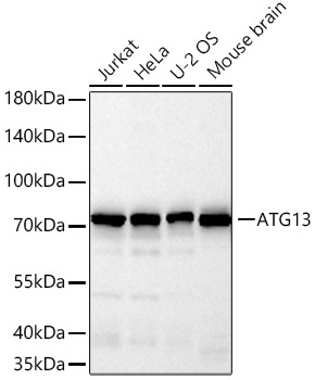
Western blot analysis of various lysates using ATG13 Rabbit pAb (CAB0690) at 1:1000 dilution incubated overnight at 4℃. Secondary antibody: HRP-conjugated Goat anti-Rabbit IgG (H+L) (CABS014) at 1:10000 dilution. Lysates/proteins: 25 μg per lane. Blocking buffer: 3% nonfat dry milk in TBST. Detection: ECL Basic Kit (AbGn00020). Exposure time: 45s.
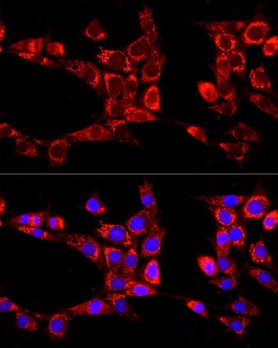
Immunofluorescence analysis of NIH/3T3 cells using ATG13 Rabbit pAb (CAB0690) at dilution of 1:100 (40x lens). Secondary antibody: Cy3-conjugated Goat anti-Rabbit IgG (H+L) (CABS007) at 1:500 dilution. Blue: DAPI for nuclear staining.