
Flow Cytometry Gating: A Comprehensive Guide
Flow Cytometry Gating: A Comprehensive Guide
Gating is how flow cytometry data is turned into meaningful results — a series of regions drawn on scatter and fluorescence plots that isolate the cells of interest while removing debris, doublets and dead cells. This guide walks through core gating strategies, from FSC/SSC and doublet discrimination to viability gating, immunophenotyping panels and FMO controls, with practical tips for clean, reproducible data.
Browse Flow Cytometry Antibodies →Contents
- Introduction to Flow Cytometry Gating
- Basic Gating Strategies
- Single-Cell Gating for Doublet Discrimination
- Dead Cell Exclusion Using Viability Dyes
- Population-Specific Gating for Immunophenotyping
- Fluorescence Minus One (FMO) Controls for Accurate Gating
- Gating Strategy Summary Table
- Practical Tips for Effective Flow Cytometry Gating
- Frequently Asked Questions
- Explore Our Antibody Range
Introduction to Flow Cytometry Gating
Gating is the process of setting criteria to identify cell populations of interest based on specific parameters in flow cytometry. Gating helps:
- Exclude unwanted events: Filters out dead cells, debris, and doublets.
- Focus on single cells: Isolates single, viable cells for precise data analysis.
- Identify specific cell populations: Differentiates based on fluorescence markers, size, and granularity.
Basic Gating Strategies
Forward Scatter (FSC) and Side Scatter (SSC) for Initial Gating
Forward Scatter (FSC) and Side Scatter (SSC) are used to analyze cell size and granularity, respectively.
- FSC: Measures cell size based on light scattered forward. Larger cells produce higher FSC values.
- SSC: Measures internal complexity or granularity. Cells with high granularity, like granulocytes, produce higher SSC values.
Setting the FSC vs. SSC Gate
- Plot FSC-A (area) vs. SSC-A: Draw a region to capture your primary cell population. This first gate will exclude debris, as debris has lower FSC and SSC values.
Single-Cell Gating for Doublet Discrimination
Doublets and clumps can resemble larger single cells, introducing variability in measurements. Doublet discrimination ensures that the analysis focuses on single cells.
Using FSC-A and FSC-H for Doublet Exclusion
- FSC-A (Area): Reflects total signal area; larger values can indicate clumps.
- FSC-H (Height): Measures the signal’s peak height, generally lower in doublets.
Plot FSC-A vs. FSC-H
- Single cells typically appear along a linear, diagonal line, while doublets deviate from this line.
- Gate along the linear population in FSC-A vs. FSC-H to exclude doublets.
FSC-W and SSC-W for Additional Doublet Gating
- FSC-W (Width): Measures pulse width, typically wider for doublets.
- Plot FSC-W vs. FSC-H: Exclude events with high FSC-W, indicative of doublets.
Dead Cell Exclusion Using Viability Dyes
Dead cells can nonspecifically bind antibodies, causing non-specific fluorescence. Use a viability dye to stain dead cells and exclude them.
Common Viability Dyes
- 7-AAD (7-Aminoactinomycin D): Enters only dead cells, showing fluorescence in dead cell populations.
- Propidium Iodide (PI): Intercalates with DNA of compromised cells, emitting fluorescence in dead cells.
- Live/Dead stains: Available in various fluorescent colors, useful in multicolor panels.
Plotting Viability
Population-Specific Gating for Immunophenotyping
Using fluorescence-conjugated antibodies, flow cytometry can differentiate cell populations based on the expression of specific markers.
Panel Design and Fluorescence Compensation
- Select non-overlapping fluorophores to minimize spectral overlap.
- Compensation controls are essential to correct for spectral overlap in multicolor experiments.
Example: T Cell Gating Strategy
For T cell analysis, specific markers like CD3 for T cells, CD4 for helper T cells, and CD8 for cytotoxic T cells are commonly used.
Step-by-Step Gating
- Set Primary FSC-A vs. SSC-A Gate: Exclude debris.
- Apply Single-Cell Gate: Use FSC-H vs. FSC-A to include single cells.
- Dead Cell Exclusion: Gate out dead cells stained with viability dyes.
- Population-Specific Gating:
Building a flow panel? Assay Genie offers conjugated antibodies and sample-prep reagents for immunophenotyping:
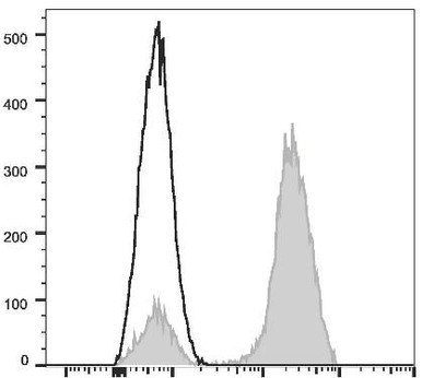
FITC Anti-Human CD3 Antibody
FITC anti-human CD3 (OKT-3), a core T-cell marker for immunophenotyping panels.
View product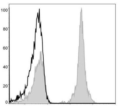
PE Anti-Human CD4 Antibody
PE anti-human CD4 (RPA-T4) to identify helper T cells by flow cytometry.
View product
Intracellular Fixation / Permeabilization Kit
Fix and permeabilise cells for intracellular flow cytometry staining.
View productFluorescence Minus One (FMO) Controls for Accurate Gating
FMO controls are critical in multicolor flow cytometry to set accurate gates, as they show fluorescence spillover from other channels without the specific marker in question. Use FMO controls to:
- Determine where to set boundaries between positive and negative populations.
- Avoid false positives due to spillover in overlapping fluorescence channels.
Gating Strategy Summary Table
Gating Step | Parameters | Purpose |
|---|---|---|
Initial FSC/SSC Gate | FSC-A vs. SSC-A | Excludes debris based on size and granularity |
Single-Cell Gate | FSC-A vs. FSC-H | Excludes doublets |
Doublet Discrimination | FSC-W vs. FSC-H | Additional doublet exclusion |
Dead Cell Exclusion | Viability Dye | Excludes dead cells |
Population-Specific Gating | Marker Fluorescence | Identifies specific cell populations |
FMO Controls | Individual Fluorophores | Sets accurate boundaries for multicolor panels |
Practical Tips for Effective Flow Cytometry Gating
- Adjust gates for each experiment: Cell populations can vary by sample, so adjust gates accordingly.
- Run controls: Include positive and negative controls to accurately identify populations.
- Use sequential gating: Move from broad gating (debris exclusion) to more specific gating (population markers).
- Optimize compensation: Use single-stain controls to set compensation correctly in multicolor
experiments.
By following these gating strategies and using controls, you can achieve precise identification and quantification of cell populations in flow cytometry, ensuring reliable and interpretable data.
Frequently Asked Questions
What is gating in flow cytometry?
Gating is drawing regions on scatter or fluorescence plots to select a specific cell population for analysis while excluding debris, doublets and dead cells.
Why is doublet discrimination important?
Doublets — two cells passing the laser together — can be mistaken for single cells with double signal, so excluding them (using FSC-A vs FSC-H) improves accuracy.
What are FMO controls?
Fluorescence-Minus-One controls contain every fluorophore in a panel except one, and are used to set accurate gates by accounting for spectral spillover.
How do you exclude dead cells?
A viability dye is added so that dead cells, which take up the dye, can be gated out before analysing the population of interest.
Explore Our Antibody Range
Assay Genie’s antibody range spans every major application and validation type — explore the category that fits your experiment:
Related reading
Designing a flow cytometry panel?
From conjugated antibodies against CD markers to fixation and permeabilization reagents, Assay Genie supplies the tools behind clean immunophenotyping — backed by expert technical support.
Browse Flow Cytometry Antibodies →Recent Posts
-
Suspected Plague Death at a Siberian Anti-Plague Institute: What Researchers Should Know
Infectious Disease · News AnalysisSuspected Plague Death at a Siberian Anti-Plague Institute: What …6th Oct 2026 -
LPS ELISA Troubleshooting: The Importance of Sample Nature
Quick answer A researcher measuring circulating lipopolysaccharide (LPS) in human serum …25th Sep 2026 -

Sekundärantikörper wählen: Die Regel, die im Labor wirklich zählt
Sekundärantikörper wählen: Die Regel, die im Labor wirklich zählt Wählen Sie einen Sekundärantikörp …24th Sep 2026