The BNIP2 Antibody (CAB6282) is a high-quality antibody developed for reliable detection and analysis of target proteins. This antibody, produced in rabbits, exhibits high reactivity with human samples and has been validated for use in Western blot applications. By specifically binding to the BNIP2 protein, researchers can accurately detect and analyze its expression in various cell types, making it a valuable asset in studies related to cell death pathways, cancer research, and cardiovascular disease.BNIP2, also known as BCL2/adenovirus E1B 19kDa interacting protein 2, is involved in regulating mitochondrial function and apoptotic pathways, making it a crucial target for understanding disease mechanisms and potential therapeutic interventions.
This antibody is validated for use in WB, ELISA applications and has demonstrated reactivity against Human, Mouse samples.
Product Name:
BNIP2 Antibody
SKU:
CAB6282
Size:
20μL, 100μL
Reactivity:
Human, Mouse
Conjugate:
Unconjugated
Immunogen:
Recombinant protein (or fragment).This information is considered to be commercially sensitive.
Recommended starting concentration is 1 μg/mL. Please optimize the concentration based on your specific assay requirements.
Synonyms:
NIP2, BNIP-2, BNIP2
Positive Sample:
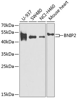
U-937, SW480, NCI-H460, Mouse heart
Cellular Localization:
Cytoplasm, Perinuclear Region.
Calculated MW:
36kDa
Observed MW:
45-55kDa
This gene is a member of the BCL2/adenovirus E1B 19 kd-interacting protein (BNIP) family. It interacts with the E1B 19 kDa protein, which protects cells from virally-induced cell death. The encoded protein also interacts with E1B 19 kDa-like sequences of BCL2, another apoptotic protector. Alternate splicing results in multiple transcript variants.
Purification Method
Affinity purification
Gene ID
663
RRID
AB_2766887
Buffer Information
Store at -20℃. Avoid freeze / thaw cycles. Buffer: PBS containing 50% glycerol, preserved with proclin300 or sodium azide, pH 7.3.
Western blot analysis of various lysates using BNIP2 Rabbit pAb (CAB6282) at 1:1000 dilution. Secondary antibody: HRP-conjugated Goat anti-Rabbit IgG (H+L) (CABS014) at 1:10000 dilution. Lysates/proteins: 25μg per lane. Blocking buffer: 3% nonfat dry milk in TBST. Detection: ECL Enhanced Kit (AbGn00021). Exposure time: 90s.