The BRMS1 Antibody (CAB14865) is a high-quality antibody developed for reliable detection and analysis of target proteins. This antibody, produced in rabbits, has high specificity and sensitivity for detecting BRMS1 protein expression in human samples, and is validated for use in Western blotting applications.BRMS1, also known as breast cancer metastasis suppressor 1, has been identified as a key player in inhibiting the spread of cancer cells to distant organs.
This antibody is validated for use in WB, IHC-P, IF/ICC, ELISA applications and has demonstrated reactivity against Human, Mouse, Rat samples.
Product Name:
BRMS1 Antibody
SKU:
CAB14865
Size:
20μL, 100μL
Reactivity:
Human, Mouse, Rat
Conjugate:
Unconjugated
Immunogen:
Recombinant protein (or fragment).This information is considered to be commercially sensitive.
Recommended starting concentration is 1 μg/mL. Please optimize the concentration based on your specific assay requirements.
Synonyms:
BRMS1
Positive Sample:
Mouse kidney, Mouse testis
Cellular Localization:
Cytoplasm, Nucleus.
Calculated MW:
28kDa
Observed MW:
28kDa
This gene reduces the metastatic potential, but not the tumorogenicity, of human breast cancer and melanoma cell lines. The protein encoded by this gene localizes primarily to the nucleus and is a component of the mSin3a family of histone deacetylase complexes (HDAC). The protein contains two coiled-coil motifs and several imperfect leucine zipper motifs. Alternative splicing results in two transcript variants encoding different isoforms.
Purification Method
Affinity purification
Gene ID
25855
RRID
AB_2761745
Buffer Information
Store at -20℃. Avoid freeze / thaw cycles. Buffer: PBS with 0.01% thimerosal,50% glycerol,pH7.3.
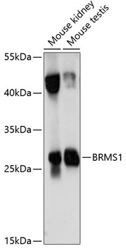
Western blot analysis of various lysates using BRMS1 Rabbit pAb (CAB14865) at 1:1000 dilution. Secondary antibody: HRP-conjugated Goat anti-Rabbit IgG (H+L) (CABS014) at 1:10000 dilution. Lysates/proteins: 25μg per lane. Blocking buffer: 3% nonfat dry milk in TBST. Detection: ECL Basic Kit (AbGn00020). Exposure time: 10s.
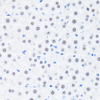
Immunohistochemistry analysis of paraffin-embedded Rat liver using BRMS1 Rabbit pAb (CAB14865) at dilution of 1:100 (40x lens). Microwave antigen retrieval performed with 0.01M PBS Buffer (pH 7.2) prior to IHC staining.
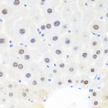
Immunohistochemistry analysis of paraffin-embedded Mouse liver using BRMS1 Rabbit pAb (CAB14865) at dilution of 1:100 (40x lens). Microwave antigen retrieval performed with 0.01M PBS Buffer (pH 7.2) prior to IHC staining.
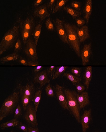
Immunofluorescence analysis of H9C2 cells using BRMS1 Rabbit pAb (CAB14865) at dilution of 1:100. Secondary antibody: Cy3-conjugated Goat anti-Rabbit IgG (H+L) (CABS007) at 1:500 dilution. Blue: DAPI for nuclear staining.