The [KO Validated] alpha-Catenin Antibody (CAB5635) is a high-quality antibody developed for reliable detection and analysis of target proteins. This antibody, produced in rabbits, demonstrates high reactivity with human samples and is validated for use in Western blot applications. By specifically binding to the Catenin Alpha 1 protein, this antibody enables precise detection and analysis in a variety of cell types, making it ideal for studies in cell biology and cancer research.Catenin Alpha 1 is a key component of the cadherin-catenin complex, which mediates cell-cell adhesion and is essential for maintaining tissue integrity and organization.
This antibody is validated for use in WB, IHC-P, IP, ELISA applications and has demonstrated reactivity against Human, Mouse, Rat samples.
Product Name:
[KO Validated] alpha-Catenin Antibody
SKU:
CAB5635
Size:
20μL, 100μL
Reactivity:
Human, Mouse, Rat
Conjugate:
Unconjugated
Immunogen:
Recombinant protein (or fragment).This information is considered to be commercially sensitive.
This gene encodes a member of the catenin family of proteins that play an important role in cell adhesion process by connecting cadherins located on the plasma membrane to the actin filaments inside the cell. The encoded mechanosensing protein contains three vinculin homology domains and undergoes conformational changes in response to cytoskeletal tension, resulting in the reconfiguration of cadherin-actin filament connections. Certain mutations in this gene cause butterfly-shaped pigment dystrophy.
Purification Method
Affinity purification
Gene ID
1495
RRID
AB_2766395
Buffer Information
Store at -20℃. Avoid freeze / thaw cycles. Buffer: PBS containing 50% glycerol, preserved with proclin300 or sodium azide, pH 7.3.
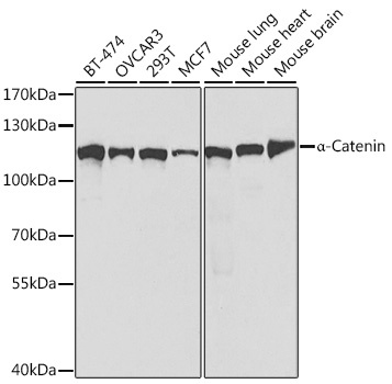
Western blot analysis of various lysates using [KO Validated] α-Catenin Rabbit pAb (CAB5635) at 1:1000 dilution. Secondary antibody: HRP-conjugated Goat anti-Rabbit IgG (H+L) (CABS014) at 1:10000 dilution. Lysates/proteins: 25μg per lane. Blocking buffer: 3% nonfat dry milk in TBST. Detection: ECL Basic Kit (AbGn00020). Exposure time: 60s.
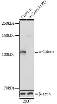
Western blot analysis of lysates from wild type (WT) and α-Catenin knockout (KO) 293T cells, using [KO Validated] α-Catenin Rabbit pAb (CAB5635) at 1:1000 dilution. Secondary antibody: HRP-conjugated Goat anti-Rabbit IgG (H+L) (CABS014) at 1:10000 dilution. Lysates/proteins: 25μg per lane. Blocking buffer: 3% nonfat dry milk in TBST. Detection: ECL Basic Kit (AbGn00020). Exposure time: 10s.
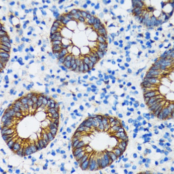
Immunohistochemistry analysis of paraffin-embedded Human vermiform appendix using [KO Validated] α-Catenin Rabbit pAb (CAB5635) at dilution of 1:100 (40x lens). Microwave antigen retrieval performed with 0.01M PBS Buffer (pH 7.2) prior to IHC staining.
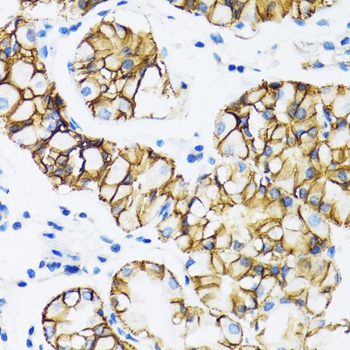
Immunohistochemistry analysis of paraffin-embedded Human stomach using [KO Validated] α-Catenin Rabbit pAb (CAB5635) at dilution of 1:100 (40x lens). Microwave antigen retrieval performed with 0.01M PBS Buffer (pH 7.2) prior to IHC staining.
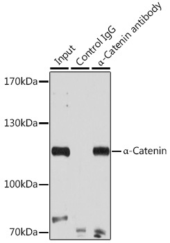
Immunoprecipitation analysis of 200 μg extracts of 293T cells using 3 μg α-Catenin antibody (CAB5635). Western blot was performed from the immunoprecipitate using α-Catenin antibody (CAB5635) at a dilution of 1:500.