The CEP43 Monoclonal Antibody (CAB21074) is a high-quality antibody developed for reliable detection and analysis of target proteins. This antibody, produced through monoclonal technology, has high specificity and sensitivity for detecting CEP43 in various biological samples. It is validated for use in immunofluorescence and immunohistochemistry applications, allowing for precise localization and visualization of CEP43 within cells.CEP43 is known to play a crucial role in centriole biogenesis and cilia assembly, making it essential for proper cell division and signaling processes.
This antibody is validated for use in WB, IF/ICC, ELISA applications and has demonstrated reactivity against Human, Mouse samples.
Product Name:
CEP43 Monoclonal Antibody
SKU:
CAB21074
Size:
20μL, 100μL
Reactivity:
Human, Mouse
Clone Number:
ARC2913
Conjugate:
Unconjugated
Immunogen:
Recombinant protein (or fragment).This information is considered to be commercially sensitive.
This gene encodes a largely hydrophilic centrosomal protein that is required for anchoring microtubules to subcellular structures. A t(6;8)(q27;p11) chromosomal translocation, fusing this gene and the fibroblast growth factor receptor 1 (FGFR1) gene, has been found in cases of myeloproliferative disorder. The resulting chimeric protein contains the N-terminal leucine-rich region of this encoded protein fused to the catalytic domain of FGFR1. Alterations in this gene may also be associated with Crohn's disease, Graves' disease, and vitiligo. Alternatively spliced transcript variants that encode different proteins have been identified.
Purification Method
Affinity purification
Gene ID
11116
Buffer Information
Store at -20℃. Avoid freeze / thaw cycles. Buffer: PBS containing 50% glycerol and 0.05% BSA, preserved with proclin300 or sodium azide, pH 7.3.
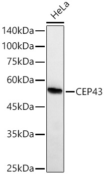
Western blot analysis of lysates from HeLa cells using CEP43 Rabbit mAb (CAB21074) at 1:1000 dilution incubated overnight at 4℃. Secondary antibody: HRP-conjugated Goat anti-Rabbit IgG (H+L) (CABS014) at 1:10000 dilution. Lysates/proteins: 25 μg per lane. Blocking buffer: 3% nonfat dry milk in TBST. Detection: ECL Basic Kit (AbGn00020). Exposure time: 90s.
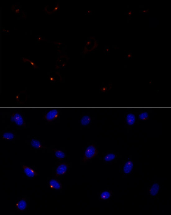
Immunofluorescence analysis of NIH/3T3 cells using CEP43 Rabbit mAb (CAB21074) at dilution of 1:50 (40x lens). Secondary antibody: Cy3-conjugated Goat anti-Rabbit IgG (H+L) (CABS007) at 1:500 dilution. Blue: DAPI for nuclear staining.
 at dilution of 1:50 (40x lens). Blue: DAPI for nuclear staining.")
 at dilution of 1:50 (40x lens). Blue: DAPI for nuclear staining.")
 at 1:10000 dilution. Lysates/proteins: 25ug per lane. Blocking buffer: 3% nonfat dry milk in TBST. Detection: ECL Basic Kit. Exposure time: 180s.")
 at 1:10000 dilution. Lysates/proteins: 25ug per lane. Blocking buffer: 3% nonfat dry milk in TBST. Detection: ECL Basic Kit. Exposure time: 90s.")
 at 1:10000 dilution. Lysates/proteins: 25ug per lane. Blocking buffer: 3% nonfat dry milk in TBST. Detection: ECL Basic Kit. Exposure time: 180s.")
 at 1:10000 dilution. Lysates/proteins: 25ug per lane. Blocking buffer: 3% nonfat dry milk in TBST. Detection: ECL Basic Kit. Exposure time: 30s.")
 at 1:10000 dilution. Lysates/proteins: 25ug per lane. Blocking buffer: 3% nonfat dry milk in TBST. Detection: ECL Basic Kit. Exposure time: 60s.")
. Perform high pressure antigen retrieval with 10 mM citrate buffer pH 6. 0 before commencing with IHC staining protocol.")
. Perform high pressure antigen retrieval with 10 mM citrate buffer pH 6. 0 before commencing with IHC staining protocol.")
 at 1:10000 dilution. Lysates/proteins: 25ug per lane. Blocking buffer: 3% nonfat dry milk in TBST. Detection: ECL Basic Kit. Exposure time: 30s.")