The CISH Antibody (CAB6286) is a high-quality antibody developed for reliable detection and analysis of target proteins. As an essential component of the suppressor of cytokine signaling (SOCS) family, CISH plays a key role in modulating immune responses and maintaining immune homeostasis. This antibody, produced in rabbits, is highly specific for human samples and is validated for use in various applications, including Western blotting. By binding to the CISH protein, researchers can accurately detect and analyze its expression in different cell types.
This antibody is validated for use in WB, ELISA applications and has demonstrated reactivity against Mouse, Rat samples.
Product Name:
CISH Antibody
SKU:
CAB6286
Size:
20μL, 100μL
Reactivity:
Mouse, Rat
Conjugate:
Unconjugated
Immunogen:
Recombinant protein (or fragment).This information is considered to be commercially sensitive.
Recommended starting concentration is 1 μg/mL. Please optimize the concentration based on your specific assay requirements.
Synonyms:
CIS, G18, SOCS, CIS-1, BACTS2, CISH
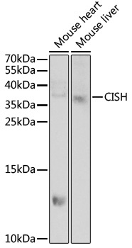
Positive Sample:
Mouse liver
Cellular Localization:
Cytosol, Plasma Membrane.
Calculated MW:
29kDa
Observed MW:
37kDa
The protein encoded by this gene contains a SH2 domain and a SOCS box domain. The protein thus belongs to the cytokine-induced STAT inhibitor (CIS), also known as suppressor of cytokine signaling (SOCS) or STAT-induced STAT inhibitor (SSI), protein family. CIS family members are known to be cytokine-inducible negative regulators of cytokine signaling. The expression of this gene can be induced by IL2, IL3, GM-CSF and EPO in hematopoietic cells. Proteasome-mediated degradation of this protein has been shown to be involved in the inactivation of the erythropoietin receptor. Multiple transcript variants encoding different isoforms have been found for this gene.
Purification Method
Affinity purification
Gene ID
1154
RRID
AB_2766891
Buffer Information
Store at -20℃. Avoid freeze / thaw cycles. Buffer: PBS containing 50% glycerol, preserved with proclin300 or sodium azide, pH 7.3.
Western blot analysis of various lysates using CISH Rabbit pAb (CAB6286) at 1:1000 dilution. Secondary antibody: HRP-conjugated Goat anti-Rabbit IgG (H+L) (CABS014) at 1:10000 dilution. Lysates/proteins: 25μg per lane. Blocking buffer: 3% nonfat dry milk in TBST. Detection: ECL Enhanced Kit (AbGn00021). Exposure time: 60s.