The COL2A1 Antibody (CAB1560) is a high-quality antibody developed for reliable detection and analysis of target proteins. This antibody, produced in rabbits, has high reactivity with human samples and is validated for use in various applications, including Western blotting.COL2A1 is involved in the formation of collagen fibrils in cartilage and other connective tissues, making it a critical target for research in musculoskeletal disorders like osteoarthritis and genetic diseases like chondrodysplasias.
This antibody is validated for use in WB, IHC-P, IF/ICC, ELISA applications and has demonstrated reactivity against Human, Mouse, Rat samples.
Product Name:
COL2A1 Antibody
SKU:
CAB1560
Size:
20μL, 100μL
Reactivity:
Human, Mouse, Rat
Conjugate:
Unconjugated
Immunogen:
This information is considered to be commercially sensitive.
Sequence:
MSAF AGLG PREK GPDP LQYM RADQ AAGG LRQH DAEV DATL KSLN NQIE SIRS PEGS RKNP ART
Tested Applications:
WBIHC-PIF/ICCELISA
Recommended Dilution:
WB
1:500 - 1:5000
IHC-P
1:1000 - 1:5000
IF/ICC
1:50 - 1:200
ELISA
Recommended starting concentration is 1 μg/mL. Please optimize the concentration based on your specific assay requirements.
Synonyms:
AOM, ANFH, SEDC, STL1, COL11A3, COL2A1
Positive Sample:
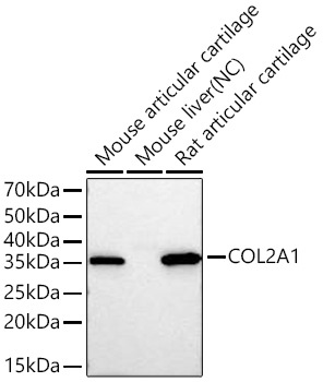
Mouse articular cartilage, Rat articular cartilage
This gene encodes the alpha-1 chain of type II collagen, a fibrillar collagen found in cartilage and the vitreous humor of the eye. Mutations in this gene are associated with achondrogenesis, chondrodysplasia, early onset familial osteoarthritis, SED congenita, Langer-Saldino achondrogenesis, Kniest dysplasia, Stickler syndrome type I, and spondyloepimetaphyseal dysplasia Strudwick type. In addition, defects in processing chondrocalcin, a calcium binding protein that is the C-propeptide of this collagen molecule, are also associated with chondrodysplasia. There are two transcripts identified for this gene.
Purification Method
Affinity purification
Gene ID
1280
RRID
AB_2763005
Buffer Information
Store at -20℃. Avoid freeze / thaw cycles. Buffer: PBS containing 50% glycerol, preserved with proclin300 or sodium azide, pH 7.3.
Western blot analysis of various lysates using COL2A1 Rabbit pAb (CAB1560) at 1:1000 dilution incubated overnight at 4℃. Secondary antibody: HRP-conjugated Goat anti-Rabbit IgG (H+L) (CABS014) at 1:10000 dilution. Lysates/proteins: 25 μg per lane. Blocking buffer: 3% nonfat dry milk in TBST. Detection: ECL Basic Kit (AbGn00020). Negative control (NC): Mouse liver. Exposure time: 20 s.
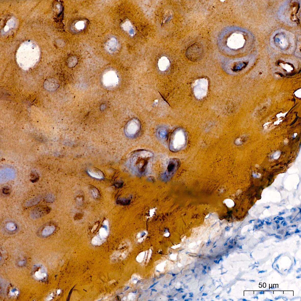
Immunohistochemistry analysis of paraffin-embedded Human cartilage tissue using COL2A1 Rabbit pAb (CAB1560) at a dilution of 1:3000 (40x lens). High pressure antigen retrieval performed with 0.01M Citrate buffer (pH 6.0) prior to IHC staining.
Vora et al.
A study of COL2A1 Expression in triple-negative breast cancer patients