The LIG1 Antibody (CAB1858) is a high-quality antibody developed for reliable detection and analysis of target proteins. This antibody, raised in rabbits, is specifically designed to target DNA Ligase 1, an essential enzyme involved in the joining of DNA strands during DNA replication and repair.With high reactivity towards human samples, this antibody is validated for use in techniques such as Western blotting, immunoprecipitation, and immunofluorescence. Its ability to specifically bind to DNA Ligase 1 enables researchers to accurately detect and analyze this crucial enzyme in a variety of cell types.DNA Ligase 1 is essential for maintaining genomic integrity and stability, making it a vital component in understanding DNA repair processes and mechanisms.
This antibody is validated for use in WB, IF/ICC, ELISA applications and has demonstrated reactivity against Human, Mouse samples.
Product Name:
LIG1 Antibody
SKU:
CAB1858
Size:
20μL, 100μL
Reactivity:
Human, Mouse
Conjugate:
Unconjugated
Immunogen:
Recombinant protein (or fragment).This information is considered to be commercially sensitive.
Recommended starting concentration is 1 μg/mL. Please optimize the concentration based on your specific assay requirements.
Synonyms:
LIGI, IMD96, hLig1, LIG1
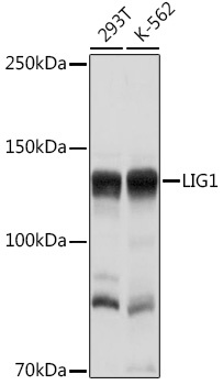
Positive Sample:
293T, K-562
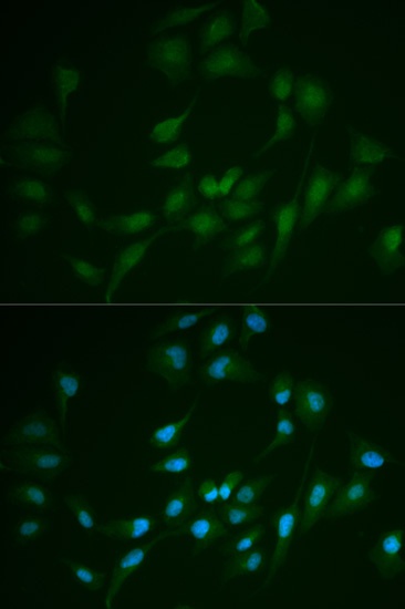
Cellular Localization:
Nucleus.
Calculated MW:
102kDa
Observed MW:
140kDa
This gene encodes a member of the ATP-dependent DNA ligase protein family. The encoded protein functions in DNA replication, recombination, and the base excision repair process. Mutations in this gene that lead to DNA ligase I deficiency result in immunodeficiency and increased sensitivity to DNA-damaging agents. Disruption of this gene may also be associated with a variety of cancers. Alternative splicing results in multiple transcript variants.
Purification Method
Affinity purification
Gene ID
3978
RRID
AB_2763893
Buffer Information
Store at -20℃. Avoid freeze / thaw cycles. Buffer: PBS containing 50% glycerol, preserved with proclin300 or sodium azide, pH 7.3.
Western blot analysis of various lysates using LIG1 Rabbit pAb (CAB1858) at 1:1000 dilution. Secondary antibody: HRP-conjugated Goat anti-Rabbit IgG (H+L) (CABS014) at 1:10000 dilution. Lysates/proteins: 25μg per lane. Blocking buffer: 3% nonfat dry milk in TBST. Detection: ECL Basic Kit (AbGn00020). Exposure time: 1s.
Immunofluorescence analysis of MCF-7 cells using LIG1 Rabbit pAb (CAB1858). Secondary antibody: Cy3-conjugated Goat anti-Rabbit IgG (H+L) (CABS007) at 1:500 dilution. Blue: DAPI for nuclear staining.