The EBP1/PA2G4 Monoclonal Antibody (CAB5114) is a high-quality antibody developed for reliable detection and analysis of target proteins. This antibody, produced in rabbits, exhibits high specificity and sensitivity towards human samples, making it an excellent choice for Western blot applications.The EBP1/PA2G4 protein is a multifunctional protein that plays a crucial role in regulating cell cycle progression and cell proliferation. It is known to interact with a variety of proteins involved in cancer development and progression, making it a potential target for cancer research.
This antibody is validated for use in WB, IHC-P, IF/ICC, ELISA applications and has demonstrated reactivity against Human, Mouse, Rat samples.
Product Name:
EBP1/PA2G4 Monoclonal Antibody
SKU:
CAB5114
Size:
20μL, 100μL
Reactivity:
Human, Mouse, Rat
Clone Number:
ARC1281
Conjugate:
Unconjugated
Immunogen:
Recombinant protein (or fragment).This information is considered to be commercially sensitive.
Recommended starting concentration is 1 μg/mL. Please optimize the concentration based on your specific assay requirements.
Synonyms:
EBP1, HG4-1, ITAF45, p38-2G4, EBP1/PA2G4
Positive Sample:
HeLa, 293T, Jurkat, Mouse lung, Mouse brain, Rat brain
Cellular Localization:
Cytoplasm, Nucleus, Nucleolus.
Calculated MW:
44kDa
Observed MW:
42KD/48kDa
This gene encodes an RNA-binding protein that is involved in growth regulation. This protein is present in pre-ribosomal ribonucleoprotein complexes and may be involved in ribosome assembly and the regulation of intermediate and late steps of rRNA processing. This protein can interact with the cytoplasmic domain of the ErbB3 receptor and may contribute to transducing growth regulatory signals. This protein is also a transcriptional co-repressor of androgen receptor-regulated genes and other cell cycle regulatory genes through its interactions with histone deacetylases. This protein has been implicated in growth inhibition and the induction of differentiation of human cancer cells. Six pseudogenes, located on chromosomes 3, 6, 9, 18, 20 and X, have been identified.
Purification Method
Affinity purification
Gene ID
5036
RRID
AB_2863451
Buffer Information
Store at -20℃. Avoid freeze / thaw cycles. Buffer: PBS containing 50% glycerol and 0.05% BSA, preserved with proclin300 or sodium azide, pH 7.3.
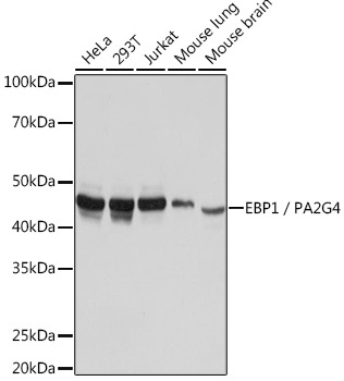
Western blot analysis of various lysates using EBP1/PA2G4 Rabbit mAb (CAB5114) at 1:1000 dilution. Secondary antibody: HRP-conjugated Goat anti-Rabbit IgG (H+L) (CABS014) at 1:10000 dilution. Lysates/proteins: 25μg per lane. Blocking buffer: 3% nonfat dry milk in TBST. Detection: ECL Basic Kit (AbGn00020). Exposure time: 3s.
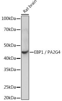
Western blot analysis of lysates from Rat brain, using EBP1/PA2G4 Rabbit mAb (CAB5114) at 1:1000 dilution. Secondary antibody: HRP-conjugated Goat anti-Rabbit IgG (H+L) (CABS014) at 1:10000 dilution. Lysates/proteins: 25μg per lane. Blocking buffer: 3% nonfat dry milk in TBST. Detection: ECL Basic Kit (AbGn00020). Exposure time: 10s.
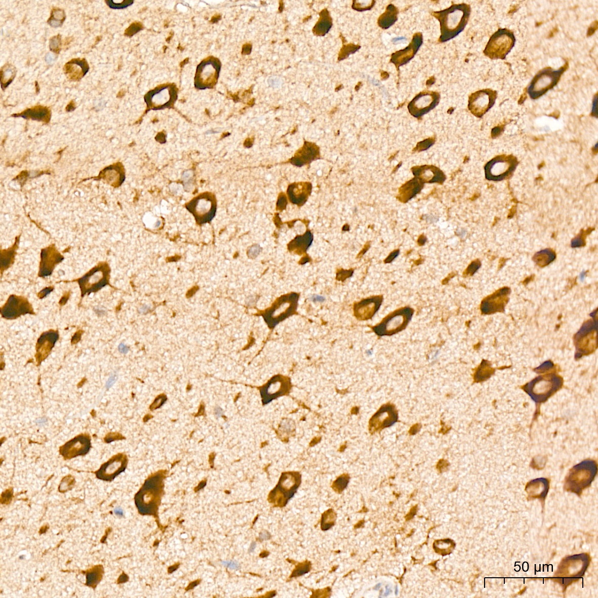
Immunohistochemistry analysis of paraffin-embedded Rat brain tissue using EBP1/PA2G4 Rabbit mAb (CAB5114) at a dilution of 1:800 (40x lens). High pressure antigen retrieval was performed with 0.01 M citrate buffer (pH 6.0) prior to IHC staining.
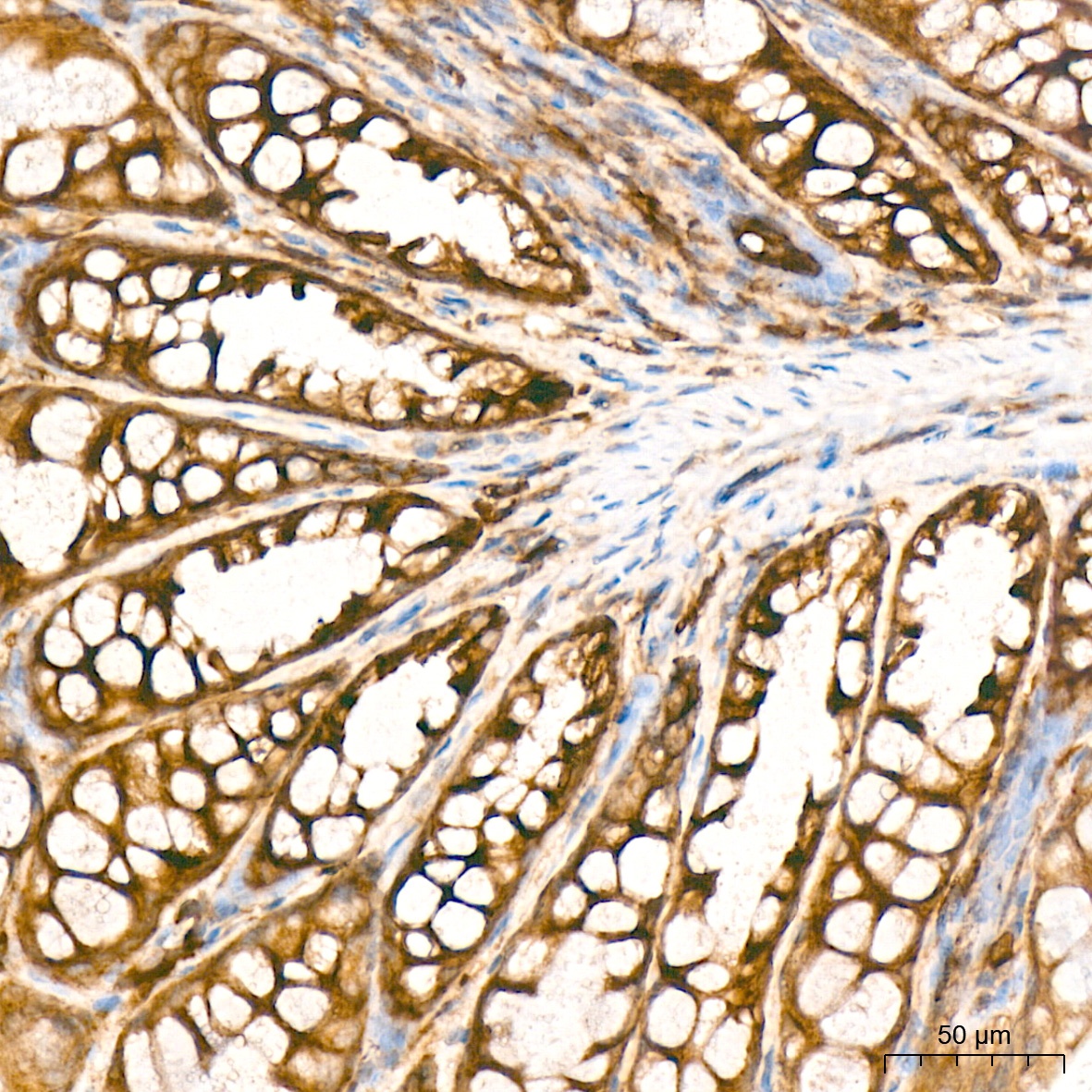
Immunohistochemistry analysis of paraffin-embedded Mouse colon tissue using EBP1/PA2G4 Rabbit mAb (CAB5114) at a dilution of 1:800 (40x lens). High pressure antigen retrieval was performed with 0.01 M citrate buffer (pH 6.0) prior to IHC staining.
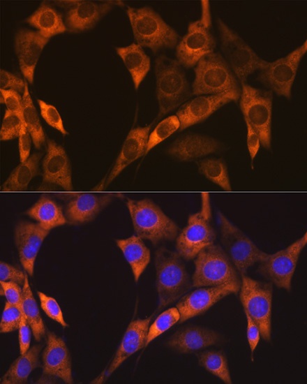
Immunofluorescence analysis of NIH-3T3 cells using EBP1/PA2G4 Rabbit mAb (CAB5114) at dilution of 1:100 (40x lens). Secondary antibody: Cy3-conjugated Goat anti-Rabbit IgG (H+L) (CABS007) at 1:500 dilution. Blue: DAPI for nuclear staining.


![Anti-EBP1 [R06-3R8] Monoclonal Antibody (AGMB00080)](https://cdn11.bigcommerce.com/s-h68l9z2lnx/images/stencil/590x590/products/271369/694985/anti-ebp1-r06-3r8-monoclonal-antibody-agmb00080__99625.1774514231.jpg?c=2 "Anti-EBP1 [R06-3R8] Monoclonal Antibody (AGMB00080)")
![Anti-EBP1 [R04-1C7] Monoclonal Antibody (AGMB02471)](https://cdn11.bigcommerce.com/s-h68l9z2lnx/images/stencil/590x590/products/273760/680104/anti-ebp1-r04-1c7-monoclonal-antibody-agmb02471__24458.1773041056.jpg?c=2 "Anti-EBP1 [R04-1C7] Monoclonal Antibody (AGMB02471)")
 (RPES4693)")