The eEF1G Antibody (CAB7891) is a high-quality antibody developed for reliable detection and analysis of target proteins. This antibody, produced in rabbits, exhibits high specificity and sensitivity towards human EEF1G samples, making it suitable for Western blot applications.EEF1G is a key component of the Eukaryotic Elongation Factor 1 complex, which plays a crucial role in delivering aminoacyl tRNAs to the ribosome during translation. By targeting EEF1G, researchers can investigate the mechanisms underlying protein synthesis and explore its implications in various cellular processes.
This antibody is validated for use in WB, IHC-P, IF/ICC, ELISA applications and has demonstrated reactivity against Human, Mouse, Rat samples.
Product Name:
eEF1G Antibody
SKU:
CAB7891
Size:
20μL, 100μL
Reactivity:
Human, Mouse, Rat
Conjugate:
Unconjugated
Immunogen:
Recombinant protein (or fragment).This information is considered to be commercially sensitive.
This gene encodes a subunit of the elongation factor-1 complex, which is responsible for the enzymatic delivery of aminoacyl tRNAs to the ribosome. This subunit contains an N-terminal glutathione transferase domain, which may be involved in regulating the assembly of multisubunit complexes containing this elongation factor and aminoacyl-tRNA synthetases.
Purification Method
Affinity purification
Gene ID
1937
RRID
AB_2769267
Buffer Information
Store at -20℃. Avoid freeze / thaw cycles. Buffer: PBS containing 50% glycerol, preserved with proclin300 or sodium azide, pH 7.3.
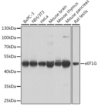
Western blot analysis of various lysates using eEF1G Rabbit pAb (CAB7891) at 1:1000 dilution. Secondary antibody: HRP-conjugated Goat anti-Rabbit IgG (H+L) (CABS014) at 1:10000 dilution. Lysates/proteins: 25μg per lane. Blocking buffer: 3% nonfat dry milk in TBST. Detection: ECL Basic Kit (AbGn00020). Exposure time: 5s.
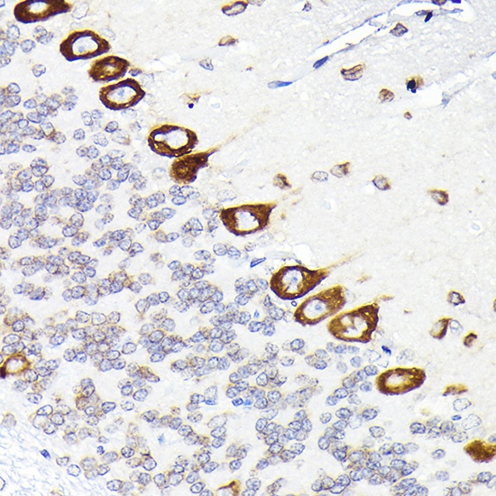
Immunohistochemistry analysis of paraffin-embedded Rat brain using eEF1G Rabbit pAb (CAB7891) at dilution of 1:50 (40x lens). High pressure antigen retrieval performed with 0.01M Citrate buffer (pH 6.0) prior to IHC staining.
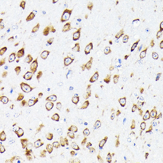
Immunohistochemistry analysis of paraffin-embedded Mouse brain using eEF1G Rabbit pAb (CAB7891) at dilution of 1:50 (40x lens). High pressure antigen retrieval performed with 0.01M Citrate buffer (pH 6.0) prior to IHC staining.
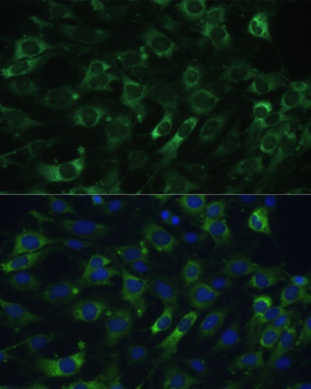
Immunofluorescence analysis of C6 cells using eEF1G Rabbit pAb (CAB7891) at dilution of 1:100 (40x lens). Secondary antibody: Cy3-conjugated Goat anti-Rabbit IgG (H+L) (CABS007) at 1:500 dilution. Blue: DAPI for nuclear staining.