The Flotillin 1 Monoclonal Antibody (CAB3023) is a high-quality antibody developed for reliable detection and analysis of target proteins. This antibody, generated in rabbits, exhibits high specificity and sensitivity towards human samples, making it suitable for use in Western blotting, immunofluorescence, and immunoprecipitation experiments.Flotillin-1 is a widely-studied protein that is implicated in diverse biological functions, including cell adhesion, migration, and intracellular signaling pathways.
This antibody is validated for use in WB, IHC-P, ELISA applications and has demonstrated reactivity against Human, Mouse, Rat samples.
Product Name:
Flotillin 1 Monoclonal Antibody
SKU:
CAB3023
Size:
20μL, 100μL
Reactivity:
Human, Mouse, Rat
Clone Number:
ARC0765
Conjugate:
Unconjugated
Immunogen:
Synthetic peptide. This information is considered to be commercially sensitive.
This gene encodes an protein that localizes to the caveolae, which are small domains on the inner cell membranes. This protein plays a role in vesicle trafficking and cell morphology. Alternative splicing results in multiple transcript variants.
Purification Method
Affinity purification
Gene ID
10211
RRID
AB_2863023
Buffer Information
Store at -20℃. Avoid freeze / thaw cycles. Buffer: PBS containing 50% glycerol and 0.05% BSA, preserved with proclin300 or sodium azide, pH 7.3.
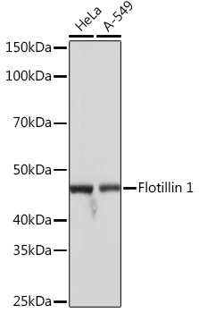
Western blot analysis of various lysates, using Flotillin 1 Rabbit mAb (CAB3023) at 1:1000 dilution incubated overnight at 4℃. Secondary antibody: HRP-conjugated Goat anti-Rabbit IgG (H+L) (CABS014) at 1:10000 dilution. Lysates/proteins: 25μg per lane. Blocking buffer: 3% nonfat dry milk in TBST. Detection: ECL Basic Kit (AbGn00020). Exposure time: 1s.
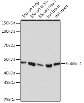
Western blot analysis of various lysates, using Flotillin 1 Rabbit mAb (CAB3023) at 1:1000 dilution incubated overnight at 4℃. Secondary antibody: HRP-conjugated Goat anti-Rabbit IgG (H+L) (CABS014) at 1:10000 dilution. Lysates/proteins: 25μg per lane. Blocking buffer: 3% nonfat dry milk in TBST. Detection: ECL Basic Kit (AbGn00020). Exposure time: 3min.
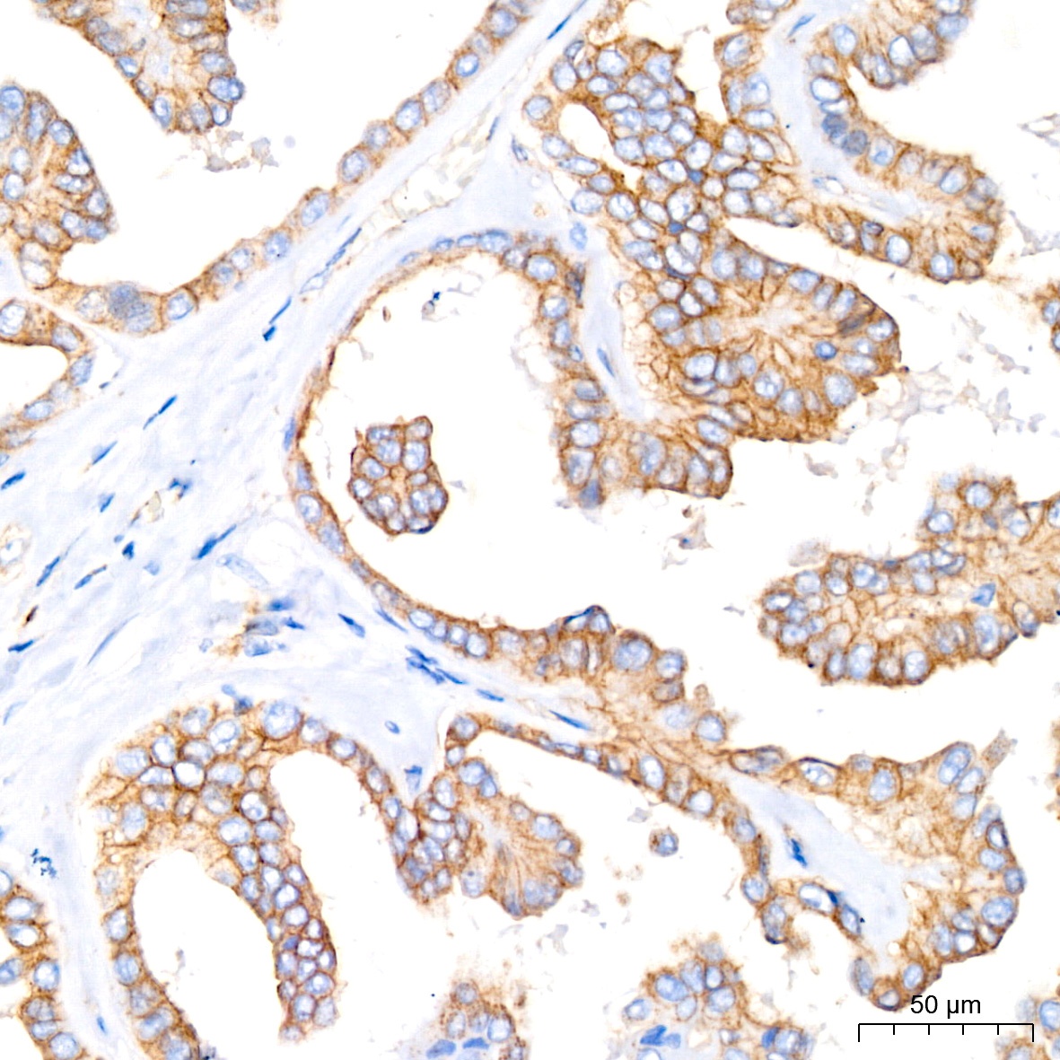
Immunohistochemistry analysis of paraffin-embedded Human thyroid cancer tissue using Flotillin 1 Rabbit mAb (CAB3023) at a dilution of 1:200 (40x lens). High pressure antigen retrieval performed with 0.01M Citrate buffer (pH 6.0) prior to IHC staining.
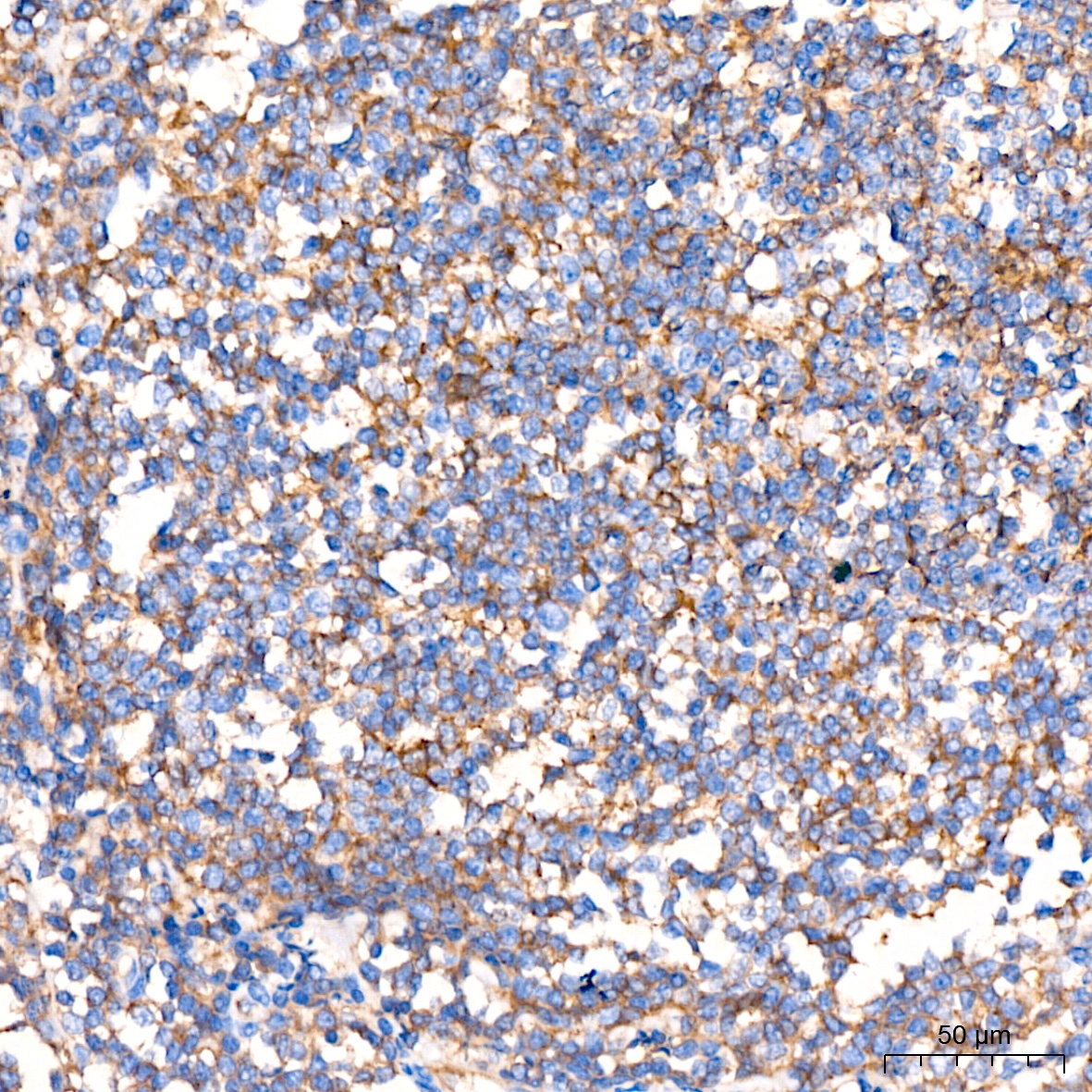
Immunohistochemistry analysis of paraffin-embedded Human tonsil tissue using Flotillin 1 Rabbit mAb (CAB3023) at a dilution of 1:200 (40x lens). High pressure antigen retrieval performed with 0.01M Citrate buffer (pH 6.0) prior to IHC staining.


![Anti-Flotillin 1 [R06-8I8] Monoclonal Antibody (AGMB01886)](https://cdn11.bigcommerce.com/s-h68l9z2lnx/images/stencil/590x590/products/273175/680118/anti-flotillin-1-r06-8i8-monoclonal-antibody-agmb01886__03314.1773041068.jpg?c=2 "Anti-Flotillin 1 [R06-8I8] Monoclonal Antibody (AGMB01886)")
![Anti-Flotillin 1 [6H9-1D9-3E10] Monoclonal Antibody (AGMB04404)](https://cdn11.bigcommerce.com/s-h68l9z2lnx/images/stencil/590x590/products/275692/677866/anti-flotillin-1-6h9-1d9-3e10-monoclonal-antibody-agmb04404__80301.1773033978.jpg?c=2 "Anti-Flotillin 1 [6H9-1D9-3E10] Monoclonal Antibody (AGMB04404)")
![Anti-Flotillin 1 [R08-5E9] Monoclonal Antibody - Knockout Validated (AGMB06641)](https://cdn11.bigcommerce.com/s-h68l9z2lnx/images/stencil/590x590/products/277922/734340/anti-flotillin-1-r08-5e9-monoclonal-antibody-knockout-validated-agmb06641__45428.1777192879.jpg?c=2 "Anti-Flotillin 1 [R08-5E9] Monoclonal Antibody - Knockout Validated (AGMB06641)")
![Anti-Flotillin 2 [R01-3I1] Monoclonal Antibody (AGMB00784)](https://cdn11.bigcommerce.com/s-h68l9z2lnx/images/stencil/590x590/products/272073/692905/anti-flotillin-2-r01-3i1-monoclonal-antibody-agmb00784__21492.1774507642.jpg?c=2 "Anti-Flotillin 2 [R01-3I1] Monoclonal Antibody (AGMB00784)")