Description
Glycolysis Assay Kit (BA0086) (BA0086)
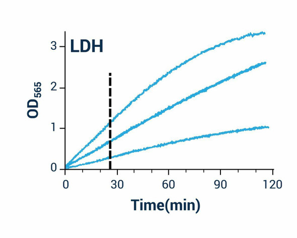
The Glycolysis Assay Kit (SKU: BA0086) provides a simple, high-throughput procedure for measuring the glycolytic rate of cells. Glycolysis is a major energy-producing metabolic pathway that generates pyruvate, one fate of which is conversion to L-lactate by lactate dehydrogenase, allowing L-lactate to serve as an indicator of glycolysis. This kit measures the production of L-lactate secreted into the cell media using a coupled reaction in which lactate dehydrogenase oxidises L-lactate to generate pyruvate and NADH, which reduces a formazan dye. The intensity of the reduced dye, measured at 565 nm, is directly proportional to the L-lactate concentration and hence the glycolytic rate of the cells.
| Product Name: | Glycolysis Assay Kit (BA0086) |
| SKU: | BA0086 |
| Detection Method: | Colorimetric |
| Detection Range: | Up to 10 mM L-lactate |
| Sample Type: | Cell culture media (adherent and suspension cells) |
| Species Reactivity: | All |
| Assay Time: | 30 minutes at room temperature |
| Kit Size: | 100 Assays |
| Equipment Required: | Microplate reader |
| Storage: | -20C |
| Shelf Life: | 6 months after receipt |
| Shipping: | Gel Pack |
Quantitative colorimetric determination of glycolysis via L-lactate. Lactate dehydrogenase oxidises L-lactate to generate NADH, which reduces a formazan dye read at 565 nm; the signal is proportional to L-lactate concentration and glycolytic rate.
- Fast and sensitive; uses 5 uL sample with a linear detection range up to 10 mM L-lactate
- Convenient single-working-reagent, room-temperature assay with no 37C heater required
- High-throughput add-mix-read format; readily automated
- Measures glycolytic rate through secreted L-lactate
- Direct assays of L-lactate produced by glycolysis in cell samples
- Screening of glycolysis inhibitors and the effect of drugs on glycolysis
Note: The below protocol is a sample protocol. Protocols are specific to each batch/lot. For the correct instructions please follow the protocol included in your kit.
| Step | Procedure |
| 1 | Sample preparation. Plate cells in the media of choice; once cells have adhered, replace with low-percentage FBS media (FBS <= 1% or serum-free). For suspension cells, seed at the desired cell number in low-percentage FBS media. Set 2 mL of media aside for making standards, add any treatments being tested, allow cells to reach the desired confluence and remove media for assay. |
| 2 | Standards. Prepare 500 uL of 10 mM premix by mixing 10 uL of the 0.5 M standard and 490 uL of the low-percentage FBS media, then dilute in centrifuge tubes as described in the dilution table. |
| 3 | Transfer 5 uL standards into separate wells of a clear flat-bottom 96-well plate and 5 uL of each sample into separate wells (running all wells at least in duplicate is recommended). |
| 4 | Prepare sufficient working reagent by mixing, per standard and sample well, 95 uL Assay Buffer, 1 uL Enzyme A, 1 uL Enzyme B and 8 uL NAD/MTT; fresh reconstitution is recommended. |
| 5 | Add 95 uL reagent to the four standards and the sample wells, tap to mix briefly and thoroughly and incubate 30 minutes at room temperature. |
| 6 | Read optical density at 565 nm (520-600 nm). |
Subtract the blank value (#4) from the standard values and plot dOD against standard concentrations to determine the slope. [L-Lactate] = (OD-sample - OD-blank) / slope (mM). Conversions: 1 mM L-lactate equals 9.01 mg/dL or 90.1 ppm. If your plate reader is not accurate to OD values above 1.0, use a modified 0, 1.5, 3, 5 mM standard curve.
| Component | Quantity | Storage |
| Assay Buffer | 12 mL | -20C |
| NAD/MTT | 1 mL | -20C |
| Standard (0.5 M L-Lactate) | 250 uL | -20C |
| Enzyme A | 120 uL | -20C |
| Enzyme B | 120 uL | -20C |