The HADHA Polyclonal Antibody (CAB22235) is a high-quality antibody developed for reliable detection and analysis of target proteins. This antibody, generated in rabbits, is highly specific for human samples and is validated for use in various applications, including Western blot and immunohistochemistry.HADHA plays a crucial role in energy production by catalyzing the oxidation of fatty acids within the mitochondria. Dysregulation of HADHA has been linked to metabolic disorders, including fatty acid oxidation disorders and mitochondrial diseases.
This antibody is validated for use in WB, IHC-P, IF/ICC, ELISA applications and has demonstrated reactivity against Human, Mouse samples.
Product Name:
HADHA Polyclonal Antibody
SKU:
CAB22235
Size:
20μL, 100μL
Reactivity:
Human, Mouse
Conjugate:
Unconjugated
Immunogen:
Recombinant protein (or fragment).This information is considered to be commercially sensitive.
Tested Applications:
WBIHC-PIF/ICCELISA
Recommended Dilution:
WB
1:500 - 1:1000
IHC-P
1:50 - 1:200
IF/ICC
1:50 - 1:200
ELISA
Recommended starting concentration is 1 μg/mL. Please optimize the concentration based on your specific assay requirements.
This gene encodes the alpha subunit of the mitochondrial trifunctional protein, which catalyzes the last three steps of mitochondrial beta-oxidation of long chain fatty acids. The mitochondrial membrane-bound heterocomplex is composed of four alpha and four beta subunits, with the alpha subunit catalyzing the 3-hydroxyacyl-CoA dehydrogenase and enoyl-CoA hydratase activities. Mutations in this gene result in trifunctional protein deficiency or LCHAD deficiency. The genes of the alpha and beta subunits of the mitochondrial trifunctional protein are located adjacent to each other in the human genome in a head-to-head orientation.
Purification Method
Affinity purification
Gene ID
3030
Buffer Information
Store at -20℃. Avoid freeze / thaw cycles. Buffer: PBS containing 50% glycerol, preserved with proclin300 or sodium azide, pH 7.3.
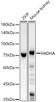
Western blot analysis of various lysates, using HADHA Rabbit pAb (CAB22235) at 1:600 dilution. Secondary antibody: HRP-conjugated Goat anti-Rabbit IgG (H+L) (CABS014) at 1:10000 dilution. Lysates/proteins: 25μg per lane. Blocking buffer: 3% nonfat dry milk in TBST. Detection: ECL Basic Kit (AbGn00020). Exposure time: 20s.
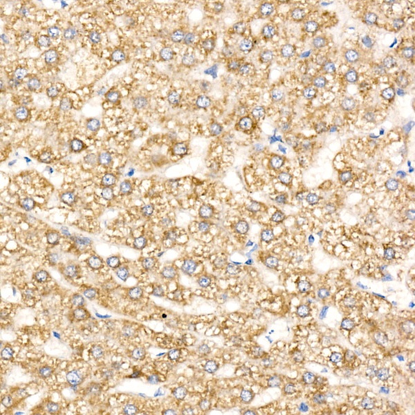
Immunohistochemistry analysis of paraffin-embedded Human liver cancer using HADHA Rabbit pAb (CAB22235) at dilution of 1:100 (40x lens). High pressure antigen retrieval performed with 0.01M Citrate buffer (pH 6.0) prior to IHC staining.
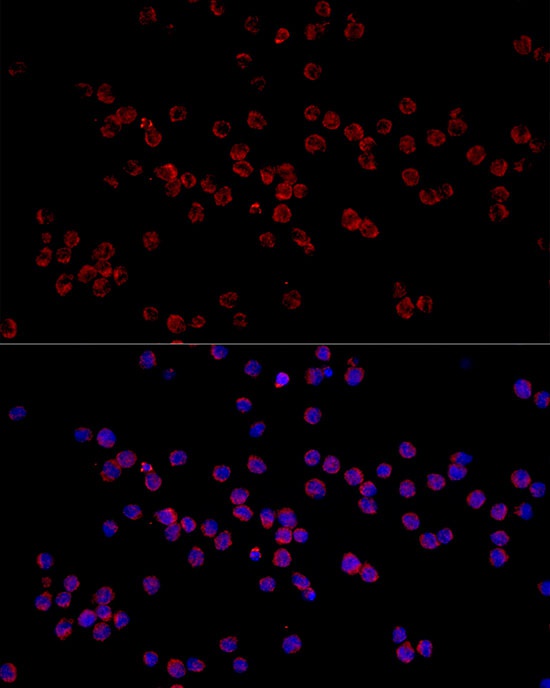
Immunofluorescence analysis of THP-1 cells using HADHA Rabbit pAb (CAB22235) at dilution of 1:50 (40x lens). Secondary antibody: Cy3-conjugated Goat anti-Rabbit IgG (H+L) (CABS007) at 1:500 dilution. Blue: DAPI for nuclear staining.
 at 1:600 dilution. Secondary antibody: HRP Goat Anti-Rabbit IgG (H+L) at 1:10000 dilution. Lysates/proteins: 25μg per lane. Blocking buffer: 3% nonfat dry milk in TBST.")
 at 1:600 dilution. Secondary antibody: HRP Goat Anti-Rabbit IgG (H+L) at 1:10000 dilution. Lysates/proteins: 25μg per lane. Blocking buffer: 3% nonfat dry milk in TBST.")

![Anti-HADHA [R07-6H4] Monoclonal Antibody (AGMB01451)](https://cdn11.bigcommerce.com/s-h68l9z2lnx/images/stencil/590x590/products/272740/680503/anti-hadha-r07-6h4-monoclonal-antibody-agmb01451__25038.1773042269.jpg?c=2 "Anti-HADHA [R07-6H4] Monoclonal Antibody (AGMB01451)")

")
")