The HGS Antibody (CAB1790) is a high-quality antibody developed for reliable detection and analysis of target proteins. This antibody, produced in rabbits, is highly reactive with human samples and has been validated for use in Western blot applications. By binding to the HGS protein, this antibody allows for easy and accurate detection and analysis in a wide range of cell types.The HGS protein, also known as Hepatocyte Growth Factor-Regulated Tyrosine Kinase Substrate, is involved in intracellular trafficking pathways and plays a crucial role in regulating cellular signaling and protein degradation.
This antibody is validated for use in WB, IF/ICC, IP, ELISA applications and has demonstrated reactivity against Human, Mouse, Rat samples.
Product Name:
HGS Antibody
SKU:
CAB1790
Size:
20μL, 100μL
Reactivity:
Human, Mouse, Rat
Conjugate:
Unconjugated
Immunogen:
Recombinant protein (or fragment).This information is considered to be commercially sensitive.
0.5μg-4μg antibody for 200μg-400μg extracts of whole cells
ELISA
Recommended starting concentration is 1 μg/mL. Please optimize the concentration based on your specific assay requirements.
Synonyms:
HRS, HGS
Positive Sample:
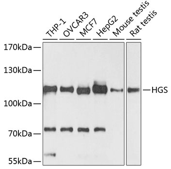
THP-1, OVCAR3, MCF7, HepG2, Mouse testis, Rat testis
Cellular Localization:
Cytoplasm, Cytoplasmic Side, Early Endosome Membrane, Endosome, Peripheral Membrane Protein, Multivesicular Body Membrane.
Calculated MW:
86kDa
Observed MW:
110kDa
The protein encoded by this gene regulates endosomal sorting and plays a critical role in the recycling and degradation of membrane receptors. The encoded protein sorts monoubiquitinated membrane proteins into the multivesicular body, targeting these proteins for lysosome-dependent degradation.
Purification Method
Affinity purification
Gene ID
9146
RRID
AB_2763831
Buffer Information
Store at -20℃. Avoid freeze / thaw cycles. Buffer: PBS with 0.01% thimerosal,50% glycerol,pH7.3.
Western blot analysis of various lysates using HGS Rabbit pAb (CAB1790) at 1:3000 dilution. Secondary antibody: HRP-conjugated Goat anti-Rabbit IgG (H+L) (CABS014) at 1:10000 dilution. Lysates/proteins: 25μg per lane. Blocking buffer: 3% nonfat dry milk in TBST. Detection: ECL Basic Kit (AbGn00020). Exposure time: 90s.
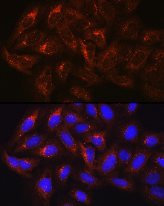
Immunofluorescence analysis of U-2 OS cells using HGS Rabbit pAb (CAB1790) at dilution of 100 (40x lens). Secondary antibody: Cy3-conjugated Goat anti-Rabbit IgG (H+L) (CABS007) at 1:500 dilution. Blue: DAPI for nuclear staining.