The Histone H3.3 Monoclonal Antibody (CAB4835) is a high-quality antibody developed for reliable detection and analysis of target proteins. This antibody, generated in rabbits, is highly specific and sensitive for the detection of histone H3.3 in human samples.Histone H3.3 is known for its unique incorporation into chromatin in a replication-independent manner, making it essential for various cellular processes such as transcriptional regulation, DNA repair, and genomic stability. The CAB4835 antibody is validated for use in Western blot applications, enabling researchers to accurately analyze histone H3.
This antibody is validated for use in WB, IHC-P, ChIP, ChIP-seq, ELISA applications and has demonstrated reactivity against Human, Mouse, Rat, Other (Wide Range Predicted) samples.
Product Name:
Histone H3.3 Monoclonal Antibody
SKU:
CAB4835
Size:
20μL, 100μL
Reactivity:
Human, Mouse, Rat, Other (Wide Range Predicted)
Clone Number:
ARC0255
Conjugate:
Unconjugated
Immunogen:
Synthetic peptide. This information is considered to be commercially sensitive.
Recommended starting concentration is 1 μg/mL. Please optimize the concentration based on your specific assay requirements.
ChIP
2μg antibody for 5μg-10μg of Chromatin
ChIP-seq
1:50 - 1:200
Synonyms:
H3F3, H3-3B, H3.3A, H3F3A, BRYLIB1, Histone H3.3
Positive Sample:
HeLa, 294T, Mouse lungRat lung
Cellular Localization:
Chromosome, Nucleus.
Calculated MW:
15kDa
Observed MW:
17kDa
Histones are basic nuclear proteins that are responsible for the nucleosome structure of the chromosomal fiber in eukaryotes. Two molecules of each of the four core histones (H2A, H2B, H3, and H4) form an octamer, around which approximately 146 bp of DNA is wrapped in repeating units, called nucleosomes. The linker histone, H1, interacts with linker DNA between nucleosomes and functions in the compaction of chromatin into higher order structures. This gene contains introns and its mRNA is polyadenylated, unlike most histone genes. The protein encoded is a replication-independent member of the histone H3 family.
Purification Method
Affinity purification
Gene ID
3020
RRID
AB_2863356
Buffer Information
Store at -20℃. Avoid freeze / thaw cycles. Buffer: PBS containing 50% glycerol and 0.05% BSA, preserved with proclin300 or sodium azide, pH 7.3.
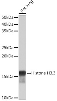
Western blot analysis of lysates from Rat lung, using Histone H3.3 Rabbit mAb (CAB4835) at 1:1000 dilution. Secondary antibody: HRP-conjugated Goat anti-Rabbit IgG (H+L) (CABS014) at 1:10000 dilution. Lysates/proteins: 25μg per lane. Blocking buffer: 3% nonfat dry milk in TBST. Detection: ECL Basic Kit (AbGn00020). Exposure time: 1s.
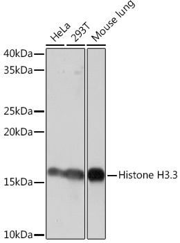
Western blot analysis of various lysates using Histone H3.3 Rabbit mAb (CAB4835) at 1:1000 dilution. Secondary antibody: HRP-conjugated Goat anti-Rabbit IgG (H+L) (CABS014) at 1:10000 dilution. Lysates/proteins: 25μg per lane. Blocking buffer: 3% nonfat dry milk in TBST. Detection: ECL Basic Kit (AbGn00020). Exposure time: 10s.
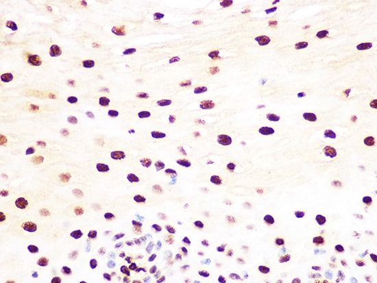
Immunohistochemistry analysis of paraffin-embedded Human esophageal using Histone H3.3 Rabbit mAb (CAB4835) at dilution of 1:100 (40x lens). Microwave antigen retrieval performed with 0.01M Tris/EDTA Buffer (pH 9.0) prior to IHC staining.
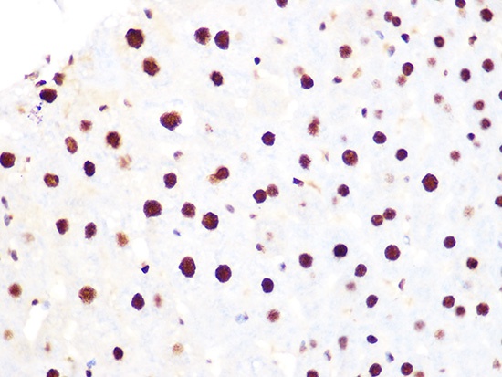
Immunohistochemistry analysis of paraffin-embedded Mouse liver using Histone H3.3 Rabbit mAb (CAB4835) at dilution of 1:100 (40x lens). Microwave antigen retrieval performed with 0.01M Tris/EDTA Buffer (pH 9.0) prior to IHC staining.
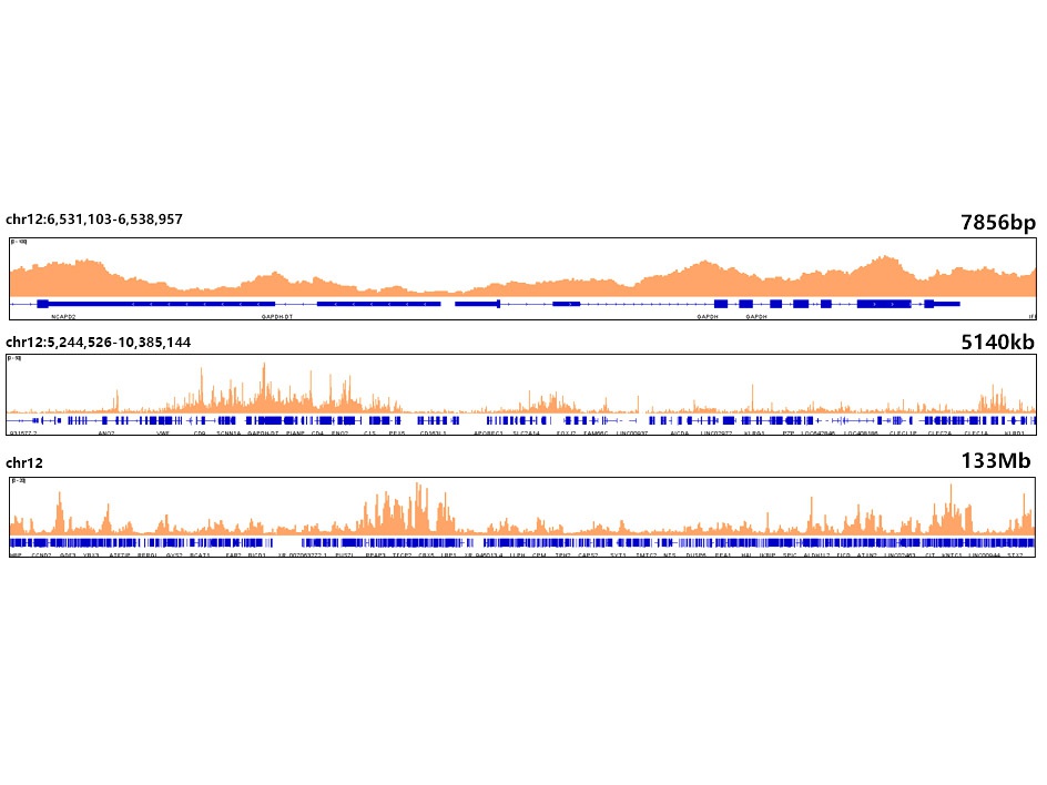
Chromatin immunoprecipitations were performed with cross-linked chromatin from HeLa cells and Histone H3.3 Rabbit mAb (CAB4835). The ChIP sequencing results indicate the enrichment pattern of Histone H3.3 in selected genomic region and representative gene loci (GAPDH), as shown in figure.


![Anti-Histone H3.3 [R04-5J3] Monoclonal Antibody (AGMB01049)](https://cdn11.bigcommerce.com/s-h68l9z2lnx/images/stencil/590x590/products/272338/692339/anti-histone-h3.3-r04-5j3-monoclonal-antibody-agmb01049__11353.1774505844.jpg?c=2 "Anti-Histone H3.3 [R04-5J3] Monoclonal Antibody (AGMB01049)")
![Anti-Phospho-Histone H3.3 (Thr3) [R21-3K-2] Monoclonal Antibody (AGMB05279)](https://cdn11.bigcommerce.com/s-h68l9z2lnx/images/stencil/590x590/products/276564/676428/anti-phospho-histone-h3.3-thr3-r21-3k-2-monoclonal-antibody-agmb05279__74671.1773029419.jpg?c=2 "Anti-Phospho-Histone H3.3 (Thr3) [R21-3K-2] Monoclonal Antibody (AGMB05279)")
