The HLA-DRA Antibody (CAB11787) is a high-quality antibody developed for reliable detection and analysis of target proteins. This antibody, produced in rabbits, is highly specific to human samples and has been validated for use in Western blot applications. By targeting the HLA-DRA protein, this antibody allows for precise detection and analysis in a variety of cell types, making it well-suited for investigations in immunology, autoimmunity, and infectious diseases.HLA-DRA is a critical component of the major histocompatibility complex (MHC) class II molecules, responsible for presenting antigen peptides to T cells and initiating adaptive immune responses.
This antibody is validated for use in WB, IHC-P, ELISA applications and has demonstrated reactivity against Human, Rat samples.
Product Name:
HLA-DRA Antibody
SKU:
CAB11787
Size:
20μL, 100μL
Reactivity:
Human, Rat
Conjugate:
Unconjugated
Immunogen:
Recombinant protein (or fragment).This information is considered to be commercially sensitive.
Recommended starting concentration is 1 μg/mL. Please optimize the concentration based on your specific assay requirements.
Synonyms:
HLA-DRA1, HLA-DRA
Positive Sample:
Raji, Rat spleen
Cellular Localization:
Cell Membrane, Endoplasmic Reticulum Membrane, Endosome Membrane, Golgi Apparatus, Late Endosome Membrane, Lysosome Membrane, Single-Pass Type I Membrane Protein, Trans-Golgi Network Membrane.
Calculated MW:
29kDa
Observed MW:
36kDa
HLA-DRA is one of the HLA class II alpha chain paralogues. This class II molecule is a heterodimer consisting of an alpha and a beta chain, both anchored in the membrane. This molecule is expressed on the surface of various antigen presenting cells such as B lymphocytes, dendritic cells, and monocytes/macrophages, and plays a central role in the immune system and response by presenting peptides derived from extracellular proteins, in particular, pathogen-derived peptides to T cells. The alpha chain is approximately 33-35 kDa and its gene contains 5 exons. Exon 1 encodes the leader peptide, exons 2 and 3 encode the two extracellular domains, and exon 4 encodes the transmembrane domain and the cytoplasmic tail. DRA does not have polymorphisms in the peptide binding part and acts as the sole alpha chain for DRB1, DRB3, DRB4 and DRB5.
Purification Method
Affinity purification
Gene ID
3122
RRID
AB_2758755
Buffer Information
Store at -20℃. Avoid freeze / thaw cycles. Buffer: PBS with 0.09% Sodium azide,50% glycerol,pH7.3.
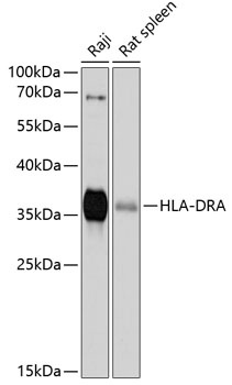
Western blot analysis of various lysates using HLA-DRA Rabbit pAb (CAB11787) at 1:1000 dilution. Secondary antibody: HRP-conjugated Goat anti-Rabbit IgG (H+L) (CABS014) at 1:10000 dilution. Lysates/proteins: 25μg per lane. Blocking buffer: 3% nonfat dry milk in TBST. Detection: ECL Basic Kit (AbGn00020). Exposure time: 90s.
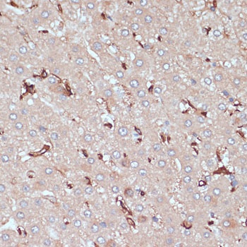
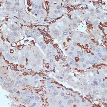
Immunohistochemistry analysis of paraffin-embedded Human liver cancer using HLA-DRA Rabbit pAb (CAB11787) at dilution of 1:100 (40x lens). Microwave antigen retrieval performed with 0.01M PBS Buffer (pH 7.2) prior to IHC staining.
Immunohistochemistry analysis of paraffin-embedded Human liver cancer using HLA-DRA Rabbit pAb (CAB11787) at dilution of 1:100 (40x lens). Microwave antigen retrieval performed with 0.01M PBS Buffer (pH 7.2) prior to IHC staining.
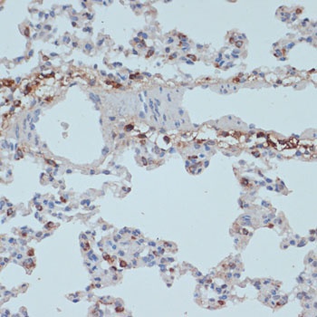
Immunohistochemistry analysis of paraffin-embedded Rat lung using HLA-DRA Rabbit pAb (CAB11787) at dilution of 1:100 (40x lens). Microwave antigen retrieval performed with 0.01M PBS Buffer (pH 7.2) prior to IHC staining.