This kit is based on a sandwich double-antibody enzyme-linked immunosorbent assay (ELISA) detection method for the quantitative measurement of Human HMGB1 in the following samples: Serum, Plasma, Cell Culture Supernatant, Cell or Tissue Lysate, Other Liquid Samples. The microplate provided in this kit has been pre-coated with a capture antibody specific to the target protein. Standards and appropriately diluted samples are added to the wells and incubated, allowing the target analyte to bind to the immobilized antibody. After incubation and washing to remove unbound components, a biotinylated detection antibody specific to a different epitope of the target protein is added, forming a sandwich complex. HRP-conjugated Streptavidin is then added, followed by TMB substrate solution to produce a colorimetric reaction. The reaction is stopped with an acidic solution, and absorbance is measured at 450 nm using a microplate reader. The signal intensity is directly proportional to the concentration of the target analyte and is determined using a standard curve.
Product Name:
Human HMGB1 ELISA Kit
SKU:
HUFI00660
Reactivity:
Human
Assay Type:
Sandwich ELISA, Double Antibody
Sensitivity:
18.75 pg/mL
Range:
31.25-2000 pg/mL
Sample Type:
Serum, Plasma, Cell Culture Supernatant, Cell or Tissue Lysate, Other Liquid Samples
Storage:
2-8°C for 12 months.
Linearity:
Sample
1:2
1:4
1:8
Serum (n = 5)
83-97%
85-100%
80-95%
EDTA Plasma (n = 5)
86-99%
82-90%
87-100%
Heparin Plasma (n = 5)
81-96%
87-101%
80-104%
Recovery:
Sample
Recovery Range (%)
Average (%)
Serum (n = 5)
85-101
97
EDTA Plasma (n = 5)
95-105
98
Heparin Plasma (n = 5)
91-103
96
Note:The below protocol is a sample protocol. Protocols are specific to each batch/lot. For the correct instructions please follow the protocol included in your kit.
Step
Procedure
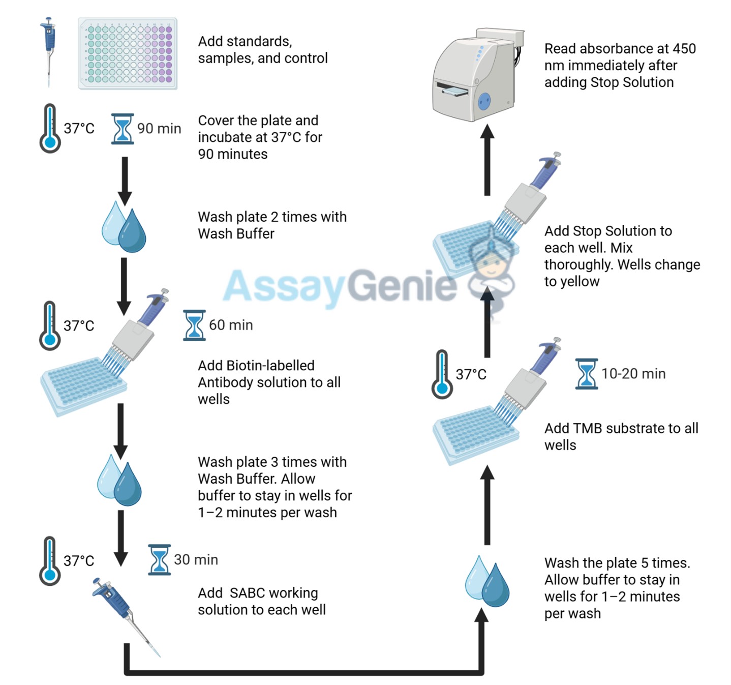
1
Reagent & Plate Preparation: Equilibrate reagents and TMB substrate to room temperature. Set standard, test sample and control (zero) wells on the pre-coated plate and record their positions.
2
Primary Incubation: Prepare standards, samples, blanks and load into designated wells. Incubate plate at 37°C for 90 minutes to allow antigen binding.
3
Detection Antibody Binding: Add biotin-labeled detection antibody and incubate at 37°C for 60 minutes.
4
HRP-Streptavidin Binding: Add HRP-Streptavidin (SABC) and incubate at 37°C for 30 minutes.
5
Color Development: Add TMB substrate and incubate in the dark for 10–20 minutes.
6
Stop Reaction & Reading: Add stop solution and measure absorbance at 450 nm immediately.
Sample Type
Protocol
Serum
Allow blood to clot, centrifuge at 1000 × g for 20 minutes, collect supernatant supernatant and store appropriately.
Plasma
Collect using anticoagulant tubes, centrifuge at 1000 × g for 15 minutes at 2–8°C and collect plasma.
Tissue Homogenate
Homogenize tissue in PBS with protease inhibitors, centrifuge and collect supernatant.
Cell Culture Supernatant
Centrifuge at 2500 rpm for 5 minutes and collect clarified supernatant.
Cell Lysate
Lyse cells using lysis buffer with protease inhibitors, centrifuge and collect protein supernatant.
Other Sample Types
For more information about how to process other sample types, (e.g., body fluids, breast milk & more), please contact our Tech Support Team at techsupport@assaygenie.com.
Component
Quantity
Storage
48T
96T
ELISA Microplate (Dismountable)
8×6
8×12
Place the test strips into a sealed foil bag with the desiccant. Store for 1 month at 2-8°C; Store for 12 months at -20°C.
Lyophilized Standard
1 vial
2 vial
Place the standards into a sealed foil bag with the desiccant. Store for 1 month at 2-8°C; Store for 12 months at -20°C.
Biotin-labeled Antibody (Concentrated, 100X)
60 ul
120 ul
2-8°C (Avoid direct light)
HRP-Streptavidin Conjugate (SABC, 100X)
60 ul
120 ul
2-8°C (Avoid direct light)
TMB Substrate
5 ml
10 ml
2-8°C (Avoid direct light)
Sample Dilution Buffer
10 ml
20 ml
2-8°C
Antibody Dilution Buffer
5 ml
10 ml
2-8°C
SABC Dilution Buffer
5 ml
10 ml
2-8°C
Stop Solution
5 ml
10 ml
2-8°C
Wash Buffer(25X)
15 ml
30 ml
2-8°C
Plate Sealer
3 pieces
5 pieces
-
Technical Manual
1 copy
1 copy
-
Nam et al.
YAP as a therapeutic target to reverse trastuzumab resistance
")
")

")
 ELISA Kit (HUEB0634)")
")
")
")
")

")