The IGF2BP3 Antibody (CAB2750) is a high-quality antibody developed for reliable detection and analysis of target proteins. This antibody, produced in rabbits, shows high reactivity with human samples and is validated for use in Western blot applications.IGF2BP3, or insulin-like growth factor 2 mRNA-binding protein 3, plays a crucial role in post-transcriptional regulation of gene expression, particularly in the context of cancer development and progression. By targeting IGF2BP3, researchers can investigate its involvement in various cellular processes, making it a versatile tool for studies in cancer biology and RNA metabolism.
This antibody is validated for use in WB, ELISA applications and has demonstrated reactivity against Human, Mouse, Rat samples.
Product Name:
IGF2BP3 Antibody
SKU:
CAB2750
Size:
20μL, 100μL
Reactivity:
Human, Mouse, Rat
Conjugate:
Unconjugated
Immunogen:
Recombinant protein (or fragment).This information is considered to be commercially sensitive.
Recommended starting concentration is 1 μg/mL. Please optimize the concentration based on your specific assay requirements.
Synonyms:
KOC, CT98, IMP3, KOC1, IMP-3, VICKZ3, IGF2BP3
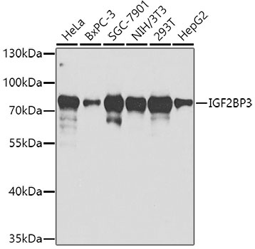
Positive Sample:
HeLa, BxPC-3, SGC-7901, NIH/3T3, 293T, HepG2
Cellular Localization:
Cytoplasm, Nucleus.
Calculated MW:
64kDa
Observed MW:
72kDa
The protein encoded by this gene is primarily found in the nucleolus, where it can bind to the 5' UTR of the insulin-like growth factor II leader 3 mRNA and may repress translation of insulin-like growth factor II during late development. The encoded protein contains several KH domains, which are important in RNA binding and are known to be involved in RNA synthesis and metabolism. A pseudogene exists on chromosome 7, and there are putative pseudogenes on other chromosomes.
Purification Method
Affinity purification
Gene ID
10643
RRID
AB_2764599
Buffer Information
Store at -20℃. Avoid freeze / thaw cycles. Buffer: PBS containing 50% glycerol, preserved with proclin300 or sodium azide, pH 7.3.
Western blot analysis of various lysates using IGF2BP3 Rabbit pAb (CAB2750) at 1:1000 dilution. Secondary antibody: HRP-conjugated Goat anti-Rabbit IgG (H+L) (CABS014) at 1:10000 dilution. Lysates/proteins: 25μg per lane. Blocking buffer: 3% nonfat dry milk in TBST. Detection: ECL Basic Kit (AbGn00020). Exposure time: 30s.