The KANK2 Antibody (CAB15420) is a high-quality antibody developed for reliable detection and analysis of target proteins. This antibody, produced in rabbits, exhibits high specificity and sensitivity when working with human samples, and has been validated for use in Western blot applications.KANK2 is known to play a critical role in various cellular processes, including cell adhesion, proliferation, and motility. Its dysregulation has been linked to cancer progression and metastasis, making it a promising target for cancer research.
This antibody is validated for use in WB, IHC-P, ELISA applications and has demonstrated reactivity against Human, Mouse, Rat samples.
Product Name:
KANK2 Antibody
SKU:
CAB15420
Size:
20μL, 100μL
Reactivity:
Human, Mouse, Rat
Conjugate:
Unconjugated
Immunogen:
Recombinant protein (or fragment).This information is considered to be commercially sensitive.
Recommended starting concentration is 1 μg/mL. Please optimize the concentration based on your specific assay requirements.
Synonyms:
SIP, MXRA3, PPKWH, NPHS16, ANKRD25, KANK2
Positive Sample:
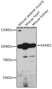
Mouse skeletal muscle, Mouse heart, Mouse kidney
Cellular Localization:
Cytoplasm, Mitochondrion.
Calculated MW:
91kDa
Observed MW:
100kDa
This gene encodes a member of the KN motif and ankyrin repeat domains (KANK) family of proteins, which play a role in cytoskeletal formation by regulating actin polymerization. The encoded protein functions in the sequestration of steroid receptor coactivators and possibly other proteins. Mutations in this gene are associated with impaired kidney podocyte function and nephrotic syndrome, and keratoderma and woolly hair.
Purification Method
Affinity purification
Gene ID
25959
RRID
AB_2762328
Buffer Information
Store at -20℃. Avoid freeze / thaw cycles. Buffer: PBS with 0.01% thimerosal,50% glycerol,pH7.3.
Western blot analysis of various lysates using KANK2 Rabbit pAb (CAB15420) at 1:1000 dilution. Secondary antibody: HRP-conjugated Goat anti-Rabbit IgG (H+L) (CABS014) at 1:10000 dilution. Lysates/proteins: 25μg per lane. Blocking buffer: 3% nonfat dry milk in TBST. Detection: ECL Basic Kit (AbGn00020). Exposure time: 180s.
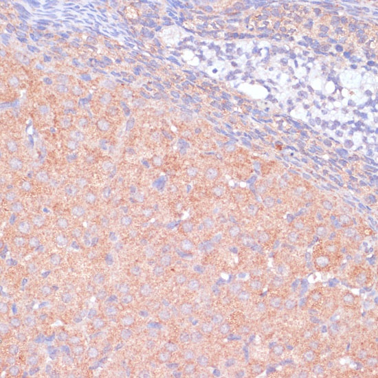
Immunohistochemistry analysis of paraffin-embedded Rat ovary using KANK2 Rabbit pAb (CAB15420) at dilution of 1:100 (40x lens). Microwave antigen retrieval performed with 0.01M PBS Buffer (pH 7.2) prior to IHC staining.