The [KO Validated] MTA2 Monoclonal Antibody (CAB4624) is a high-quality antibody developed for reliable detection and analysis of target proteins. This antibody, validated for use in various applications including Western blot and immunohistochemistry, specifically targets the MTA2 protein in human samples.MTA2 is known for its role in chromatin remodeling and gene regulation, making it a valuable target for studies in cancer research and epigenetics. By detecting and analyzing MTA2 expression in different cell types, researchers can gain insights into the mechanisms underlying gene silencing and transcriptional regulation, which are critical for understanding cancer progression and drug resistance.
This antibody is validated for use in WB, IHC-P, IF/ICC, ChIP, ELISA applications and has demonstrated reactivity against Human samples.
Product Name:
[KO Validated] MTA2 Monoclonal Antibody
SKU:
CAB4624
Size:
20μL, 100μL
Reactivity:
Human
Clone Number:
ARC1056
Conjugate:
Unconjugated
Immunogen:
Synthetic peptide. This information is considered to be commercially sensitive.
Sequence:
GVPF SANG RPLA SGIR SSSQ PAAK RQKL NPAD APNP VVFV ATKD TRAL RKAL THLE MRRA ARRP NLPL KVKP TLIA VRPP VPLP APSH PAST NEPI VLED
Tested Applications:
WBIHC-PIF/ICCChIPELISA
Recommended Dilution:
WB
1:500 - 1:1000
IHC-P
1:50 - 1:200
IF/ICC
1:50 - 1:200
ELISA
Recommended starting concentration is 1 μg/mL. Please optimize the concentration based on your specific assay requirements.
ChIP
5μg antibody for 10μg-15μg of Chromatin
Synonyms:
PID, MTA1L1, A2
Positive Sample:
HeLa, 293T
Cellular Localization:
Nucleus.
Calculated MW:
75kDa
Observed MW:
75kDa
This gene encodes a protein that has been identified as a component of NuRD, a nucleosome remodeling deacetylase complex identified in the nucleus of human cells. It shows a very broad expression pattern and is strongly expressed in many tissues. It may represent one member of a small gene family that encode different but related proteins involved either directly or indirectly in transcriptional regulation. Their indirect effects on transcriptional regulation may include chromatin remodeling. It is closely related to another member of this family, a protein that has been correlated with the metastatic potential of certain carcinomas. These two proteins are so closely related that they share the same types of domains. These domains include two DNA binding domains, a dimerization domain, and a domain commonly found in proteins that methylate DNA. One of the proteins known to be a target protein for this gene product is p53. Deacetylation of p53 is correlated with a loss of growth inhibition in transformed cells supporting a connection between these gene family members and metastasis.
Purification Method
Affinity purification
Gene ID
9219
RRID
AB_2863312
Buffer Information
Store at -20℃. Avoid freeze / thaw cycles. Buffer: PBS containing 50% glycerol and 0.05% BSA, preserved with proclin300 or sodium azide, pH 7.3.
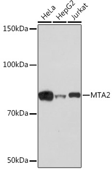
Western blot analysis of various lysates using [KO Validated] MTA2 Rabbit mAb (CAB4624) at 1:1000 dilution. Secondary antibody: HRP-conjugated Goat anti-Rabbit IgG (H+L) (CABS014) at 1:10000 dilution. Lysates/proteins: 25μg per lane. Blocking buffer: 3% nonfat dry milk in TBST. Detection: ECL Basic Kit (AbGn00020). Exposure time: 3min.
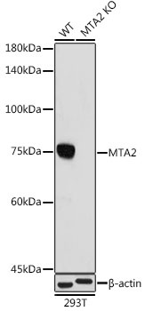
Western blot analysis of lysates from wild type (WT) and MTA2 knockout (KO) 293T cells,using [KO Validated] MTA2 Rabbit mAb (CAB4624) at 1:1000 dilution. Secondary antibody: HRP-conjugated Goat anti-Rabbit IgG (H+L) (CABS014) at 1:10000 dilution. Lysates/proteins: 25μg per lane. Blocking buffer: 3% nonfat dry milk in TBST. Detection: ECL Basic Kit (AbGn00020). Exposure time: 180s.
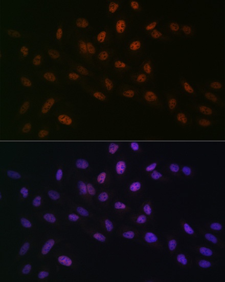
Immunofluorescence analysis of U-2 OS cells using [KO Validated] MTA2 Rabbit mAb (CAB4624) at dilution of 1 : 100 (40x lens). Secondary antibody: Cy3-conjugated Goat anti-Rabbit IgG (H+L) (CABS007) at 1:500 dilution. Blue: DAPI for nuclear staining.
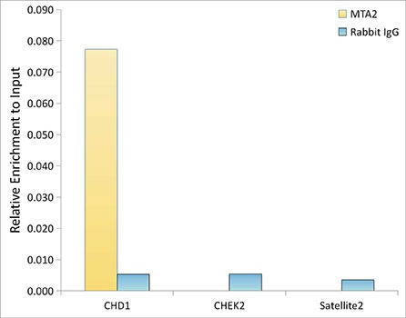
Chromatin immunoprecipitation analysis of extracts of HeLa cells, using [KO Validated] MTA2 Rabbit mAb (CAB4624) and rabbit IgG.The amount of immunoprecipitated DNA was checked by quantitative PCR. Histogram was constructed by the ratios of the immunoprecipitated DNA to the input.