The Cytokeratin 5 (KRT5) Antibody (CAB2662) is a high-quality antibody developed for reliable detection and analysis of target proteins. This antibody, produced in rabbits, exhibits high reactivity with human samples and has been validated for use in Western blot applications. It specifically targets the KRT5 protein, allowing for accurate detection and analysis in a variety of cell types.KRT5 plays a crucial role in maintaining the structural integrity of epithelial tissues and is commonly used as a marker for identifying basal cells in skin, hair follicles, and other epithelial structures.
This antibody is validated for use in WB, IHC-P, IF/ICC, IP, ELISA applications and has demonstrated reactivity against Human, Mouse samples.
Product Name:
Cytokeratin 5 (KRT5) Antibody
SKU:
CAB2662
Size:
20μL, 100μL
Reactivity:
Human, Mouse
Conjugate:
Unconjugated
Immunogen:
Synthetic peptide. This information is considered to be commercially sensitive.
The protein encoded by this gene is a member of the keratin gene family. The type II cytokeratins consist of basic or neutral proteins which are arranged in pairs of heterotypic keratin chains coexpressed during differentiation of simple and stratified epithelial tissues. This type II cytokeratin is specifically expressed in the basal layer of the epidermis with family member KRT14. Mutations in these genes have been associated with a complex of diseases termed epidermolysis bullosa simplex. The type II cytokeratins are clustered in a region of chromosome 12q12-q13.
Purification Method
Affinity purification
Gene ID
3852
RRID
AB_2764528
Buffer Information
Store at -20℃. Avoid freeze / thaw cycles. Buffer: PBS with 0.09% Sodium azide,50% glycerol,pH7.3.
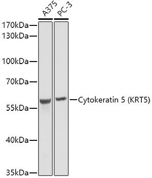
Western blot analysis of various lysates using Cytokeratin 5 Rabbit pAb (CAB2662) at 1:1000 dilution. Secondary antibody: HRP-conjugated Goat anti-Rabbit IgG (H+L) (CABS014) at 1:10000 dilution. Lysates/proteins: 25μg per lane. Blocking buffer: 3% nonfat dry milk in TBST. Detection: ECL Basic Kit (AbGn00020). Exposure time: 1s.
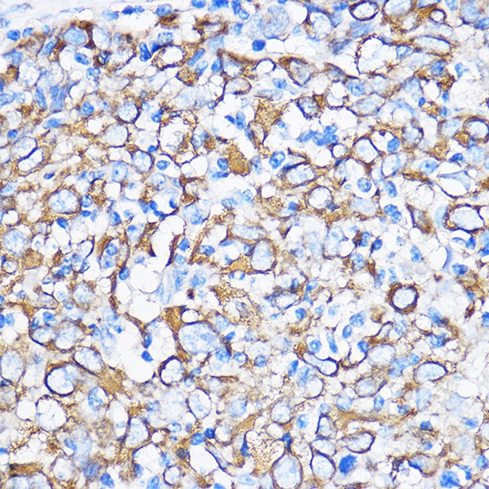
Immunohistochemistry analysis of paraffin-embedded Human esophageal cancer using Cytokeratin 5 Rabbit pAb (CAB2662) at dilution of 1:100 (40x lens). Microwave antigen retrieval performed with 0.01M PBS Buffer (pH 7.2) prior to IHC staining.
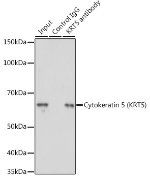
Immunoprecipitation analysis of 200 μg extracts of A-431 cells using 3 μg Cytokeratin 5 antibody (CAB2662). Western blot was performed from the immunoprecipitate using Cytokeratin 5 antibody (CAB2662) at a dilution of 1:5000.