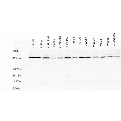
Kv1.2 Antibody [C25] is a rabbit recombinant monoclonal antibody directed against Kv1.2, offered by Assay Genie for research applications. Reported applications include WB, IHC and ICC/IF. Reported reactivity: Human, Mouse and Rat. Supplied as IgG Kappa, purified by AmMag™ Ultra AT Protein A MagBeads.
Kv1.2, encoded by the KCNA2 gene, is a voltage-gated potassium channel subunit that plays a critical role in regulating neuronal excitability and synaptic transmission. Beyond its significance as a biophysical model for ion channel gating, Kv1.2 has emerged as a key modulator of brain function, with growing evidence linking KCNA2 mutations to a range of neurological disorders, including epilepsy, ataxia, and neurodevelopmental syndromes. A landmark crystallographic study resolved the Kv1.2 channel structure at 2.9 Å, revealing the open activation gate and tetrameric subunit architecture. This structural insight has become foundational for understanding the gating mechanisms of voltage-gated ion channels across the nervous system. Functionally, Kv1.2 channels are expressed in axons and presynaptic terminals, where they shape action potential propagation and modulate neurotransmitter release. By influencing both the timing and strength of synaptic communication, Kv1.2 contributes to the fine-tuning of neural circuit dynamics essential for cognition, motor control, and sensory processing. In the context of neurodegenerative disease, dysregulation of Kv1.2 activity may lead to aberrant neuronal firing, excitotoxicity, and impaired network stability—hallmarks of conditions such as Alzheimer’s disease and multiple sclerosis. As such, Kv1.2 is increasingly recognized as a potential therapeutic target for restoring electrical balance in diseased neural circuits. Due to its recombinant nature, our Human Kv1.2 Recombinant Monoclonal IgG offers high specificity and reproducibility, making it ideal for applications in immunohistochemistry, western blotting, and ion channel research. This antibody is also available conjugated to ATTO 390, ATTO 488, ATTO 594, APC, Biotin, FITC, HRP, PerCP and RPE. Supplied in 100 µg. For research use only; not for diagnostic or therapeutic procedures.
Product Name:
Kv1.2 Antibody [C25]
Product SKU:
STMA00232
Size:
100 µg
Target:
Kv1.2
Host Species:
Rabbit
Antibody Type:
Recombinant Monoclonal
Clone ID:
C25
Research Areas:
Neuroscience, Ion Channels, Potassium Channels, Voltage-Gated Potassium Channels
Buffer:
PBS pH 7.4, 0.02% Proclin 300, 50% glycerol
Storage & Shipping:
Store at -20°C. Shipped on Blue Ice or 4°C. Conjugated variants should be stored according to the product label.
Format:
Liquid
Applications:
WB, IHC, ICC/IF
Antibody Isotype:
IgG Kappa
Purification:
AmMag™ Ultra AT Protein A MagBeads
Concentration:
1mg/mL
Reactivity:
Human, Mouse, Rat
Recommended Dilution:
WB (1:1000), IHC (1:100), ICC/IF (1:100); optimal dilutions for assays should be determined by the user.
Specificity:
Detects the cytoplasmic C-terminus of Kv1.2. Does not cross-react with other Kv1 channels.
Guarantee:
12 months from date of dispatch
Immunogen:
Fusion protein, AA #428-499 (cytoplasmic C-terminus) of human Kv1.2. Epitope mapped to within AA #463-480 (EGVNNSNEDFREENLKTA).
Brain cortex, paranodal and juxtanodal zones in myelinated spinal cord, and peroneal nerve in the juxtaparanodal regions of the nodes of Ranvier.
Alternative Names:
Kv1.2, KCNA2, Potassium voltage-gated channel subfamily A member 2, Voltage-gated potassium channel subunit Kv1.2, NGK1, HBK5, HUKIV, MK2, RBK2, Kca12
References:
1. Baronas, V. A., Yang, R. Y., & Kurata, H. T. (2017). Extracellular redox sensitivity of Kv1.2 potassium channels. Scientific Reports, 7, 9145. https://doi.org/10.1038/s41598-017-08718-z 2. Long, S. B., Campbell, E. B., & MacKinnon, R. (2005). Crystal structure of a mammalian voltage-dependent Shaker family K+ channel. Science, 309(5736), 897–903. DOI: 10.1126/science.1116269 3. Kole, M. H. P., Letzkus, J. J., & Stuart, G. J. (2007). Axon initial segment Kv1 channels control axonal action potential waveform and synaptic efficacy. Neuron, 55(4), 633–647. DOI: 10.1016/j.neuron.2007.07.031

![Western blot analysis with Rabbit Anti-Kv1.2 Monoclonal Antibody, Clone C25 (STMA00232) showing detection of Human Kv1.2 protein in a transient over-expression (OE) lysate (OriGene LY401550). Block: 2% skim milk + 2% BSA for 1 hour at RT. Primary Antibody: Rabbit Anti-Human Kv1.2 Recombinant Monoclonal [C25] (STMA00232) at 1:10000 for 2 hours at RT. Secondary Antibody: Goat anti-Rabbit IgG-HRP (H&L) at 1:5000 for 1 hour at RT.](https://cdn11.bigcommerce.com/s-h68l9z2lnx/images/stencil/1280x1280/products/279998/767904/anti-kv1.2-antibody-c25__57216.1785532762.jpg?c=2?imbypass=on "Western blot analysis with Rabbit Anti-Kv1.2 Monoclonal Antibody, Clone C25 (STMA00232) showing detection of Human Kv1.2 protein in a transient over-expression (OE) lysate (OriGene LY401550). Block: 2% skim milk + 2% BSA for 1 hour at RT. Primary Antibody: Rabbit Anti-Human Kv1.2 Recombinant Monoclonal [C25] (STMA00232) at 1:10000 for 2 hours at RT. Secondary Antibody: Goat anti-Rabbit IgG-HRP (H&L) at 1:5000 for 1 hour at RT.")
![Immunocytochemistry/Immunofluorescence analysis with Rabbit Anti-Kv1.2 Monoclonal Antibody, Clone C25 (STMA00232). Tissue: Pheochromocytoma cells (PC-12). Species: Rat. Fixation: 4% PFA for 10 min at RT. Permeabilization: 0.15% TritonX for 15 min at RT. Blocking: 10% goat serum for 40 min at RT. Primary Antibody: Rabbit Anti-Human Kv1.2 Recombinant Monoclonal [C25] (STMA00232) at 1:100 for 1 hour at RT.](https://cdn11.bigcommerce.com/s-h68l9z2lnx/images/stencil/1280x1280/products/279998/775096/anti-kv1.2-antibody-c25__20109.1785555459.jpg?c=2?imbypass=on "Immunocytochemistry/Immunofluorescence analysis with Rabbit Anti-Kv1.2 Monoclonal Antibody, Clone C25 (STMA00232). Tissue: Pheochromocytoma cells (PC-12). Species: Rat. Fixation: 4% PFA for 10 min at RT. Permeabilization: 0.15% TritonX for 15 min at RT. Blocking: 10% goat serum for 40 min at RT. Primary Antibody: Rabbit Anti-Human Kv1.2 Recombinant Monoclonal [C25] (STMA00232) at 1:100 for 1 hour at RT.")
![Immunohistochemistry analysis with Rabbit Anti-Kv1.2 Monoclonal Antibody, Clone C25 (STMA00232). Tissue: Brain. Species: Mouse. Fixation: Paraffin embedded, 4um thin. Blocking: 3% milk for 1 hour at RT. Primary Antibody: Rabbit Anti-Human Kv1.2 Recombinant Monoclonal [C25] (STMA00232) at 1:100 for 1 hour at RT. Secondary Antibody: Goat anti-Rabbit IgG-HRP (H&L) at 1:100 for 45 min at RT.](https://cdn11.bigcommerce.com/s-h68l9z2lnx/images/stencil/1280x1280/products/279998/778390/anti-kv1.2-antibody-c25__76282.1785565885.jpg?c=2?imbypass=on "Immunohistochemistry analysis with Rabbit Anti-Kv1.2 Monoclonal Antibody, Clone C25 (STMA00232). Tissue: Brain. Species: Mouse. Fixation: Paraffin embedded, 4um thin. Blocking: 3% milk for 1 hour at RT. Primary Antibody: Rabbit Anti-Human Kv1.2 Recombinant Monoclonal [C25] (STMA00232) at 1:100 for 1 hour at RT. Secondary Antibody: Goat anti-Rabbit IgG-HRP (H&L) at 1:100 for 45 min at RT.")
![Immunocytochemistry/Immunofluorescence analysis with Rabbit Anti-Kv1.2 Monoclonal Antibody, Clone C25 (STMA00232). Tissue: Neuroblastoma cells (Kelly). Species: Human. Fixation: 4% PFA for 10 min at RT. Permeabilization: 0.15% TritonX for 15 min at RT. Blocking: 10% goat serum for 40 min at RT. Primary Antibody: Rabbit Anti-Human Kv1.2 Recombinant Monoclonal [C25] (STMA00232) at 1:100 for 1 hour at RT.](https://cdn11.bigcommerce.com/s-h68l9z2lnx/images/stencil/1280x1280/products/279998/773504/anti-kv1.2-antibody-c25__79507.1785550422.jpg?c=2?imbypass=on "Immunocytochemistry/Immunofluorescence analysis with Rabbit Anti-Kv1.2 Monoclonal Antibody, Clone C25 (STMA00232). Tissue: Neuroblastoma cells (Kelly). Species: Human. Fixation: 4% PFA for 10 min at RT. Permeabilization: 0.15% TritonX for 15 min at RT. Blocking: 10% goat serum for 40 min at RT. Primary Antibody: Rabbit Anti-Human Kv1.2 Recombinant Monoclonal [C25] (STMA00232) at 1:100 for 1 hour at RT.")
![Western blot analysis with Rabbit Anti-Kv1.2 Monoclonal Antibody, Clone C25 (STMA00232) showing detection of Rat Kv1.2 protein in a Rat brain membrane lysate. Block: 2% skim milk + 2% BSA for 1 hour at RT. Primary Antibody: Rabbit Anti-Human Kv1.2 Recombinant Monoclonal [C25] (STMA00232) at 1:1000 for 2 hours at RT. Secondary Antibody: Goat anti-Rabbit IgG-HRP (H&L) at 1:5000 for 1 hour at RT.](https://cdn11.bigcommerce.com/s-h68l9z2lnx/images/stencil/1280x1280/products/279998/777167/anti-kv1.2-antibody-c25__04262.1785562039.jpg?c=2?imbypass=on "Western blot analysis with Rabbit Anti-Kv1.2 Monoclonal Antibody, Clone C25 (STMA00232) showing detection of Rat Kv1.2 protein in a Rat brain membrane lysate. Block: 2% skim milk + 2% BSA for 1 hour at RT. Primary Antibody: Rabbit Anti-Human Kv1.2 Recombinant Monoclonal [C25] (STMA00232) at 1:1000 for 2 hours at RT. Secondary Antibody: Goat anti-Rabbit IgG-HRP (H&L) at 1:5000 for 1 hour at RT.")
![Western blot analysis with Rabbit Anti-Kv1.2 Monoclonal Antibody, Clone C25 (STMA00232) showing detection of Human Kv1.2 protein in a transient over-expression (OE) lysate (OriGene LY401550). Block: 2% skim milk + 2% BSA for 1 hour at RT. Primary Antibody: Rabbit Anti-Human Kv1.2 Recombinant Monoclonal [C25] (STMA00232) at 1:10000 for 2 hours at RT. Secondary Antibody: Goat anti-Rabbit IgG-HRP (H&L) at 1:5000 for 1 hour at RT.](https://cdn11.bigcommerce.com/s-h68l9z2lnx/images/stencil/608x608/products/279998/767904/anti-kv1.2-antibody-c25__57216.1785532762.jpg?c=2 "Western blot analysis with Rabbit Anti-Kv1.2 Monoclonal Antibody, Clone C25 (STMA00232) showing detection of Human Kv1.2 protein in a transient over-expression (OE) lysate (OriGene LY401550). Block: 2% skim milk + 2% BSA for 1 hour at RT. Primary Antibody: Rabbit Anti-Human Kv1.2 Recombinant Monoclonal [C25] (STMA00232) at 1:10000 for 2 hours at RT. Secondary Antibody: Goat anti-Rabbit IgG-HRP (H&L) at 1:5000 for 1 hour at RT.")