The MYH7/beta-MHC Antibody (CAB7564) is a high-quality antibody developed for reliable detection and analysis of target proteins. This antibody, generated in rabbits, demonstrates high reactivity with human samples and has been validated for use in Western blot applications. By specifically targeting the myosin-7 protein, this antibody allows for accurate detection and analysis in a variety of cell types, making it an ideal choice for studies in cell biology, muscle physiology, and related fields.Myosin-7 is known for its role in various cellular processes, including cell migration, adhesion, and organelle transport. Dysregulation of myosin-7 has been linked to a range of diseases and disorders, including hearing loss, ciliopathies, and certain types of cancer.
This antibody is validated for use in WB, IF/ICC, ELISA applications and has demonstrated reactivity against Human, Mouse, Rat samples.
Product Name:
MYH7/beta-MHC Antibody
SKU:
CAB7564
Size:
20μL, 100μL
Reactivity:
Human, Mouse, Rat
Conjugate:
Unconjugated
Immunogen:
Synthetic peptide. This information is considered to be commercially sensitive.
Muscle myosin is a hexameric protein containing 2 heavy chain subunits, 2 alkali light chain subunits, and 2 regulatory light chain subunits. This gene encodes the beta (or slow) heavy chain subunit of cardiac myosin. It is expressed predominantly in normal human ventricle. It is also expressed in skeletal muscle tissues rich in slow-twitch type I muscle fibers. Changes in the relative abundance of this protein and the alpha (or fast) heavy subunit of cardiac myosin correlate with the contractile velocity of cardiac muscle. Its expression is also altered during thyroid hormone depletion and hemodynamic overloading. Mutations in this gene are associated with familial hypertrophic cardiomyopathy, myosin storage myopathy, dilated cardiomyopathy, and Laing distal myopathy.
Purification Method
Affinity purification
Gene ID
4625
RRID
AB_2768089
Buffer Information
Store at -20℃. Avoid freeze / thaw cycles. Buffer: PBS containing 50% glycerol, preserved with proclin300 or sodium azide, pH 7.3.
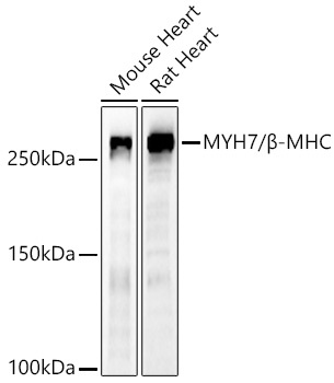
Western blot analysis of various lysates using MYH7/β-MHC Rabbit pAb (CAB7564) at 1:500 dilution. Secondary antibody: HRP-conjugated Goat anti-Rabbit IgG (H+L) (CABS014) at 1:10000 dilution. Lysates / proteins: 25 μg per lane. Blocking buffer: 3 % nonfat dry milk in TBST. Detection: ECL Basic Kit (AbGn00020). Exposure time: 180s.
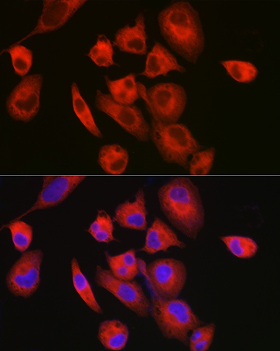
Immunofluorescence analysis of RD cells using MYH7/β-MHC Rabbit pAb (CAB7564) at dilution of 1:100 (40x lens). Secondary antibody: Cy3-conjugated Goat anti-Rabbit IgG (H+L) (CABS007) at 1:500 dilution. Blue: DAPI for nuclear staining.