The Nanog Monoclonal Antibody (CAB22625) is a high-quality antibody developed for reliable detection and analysis of target proteins. This monoclonal antibody, produced using hybridoma technology, exhibits high specificity and sensitivity in detecting Nanog expression in various tissues and cell types. Nanog is essential for maintaining stem cell identity and has implications in developmental biology, regenerative medicine, and cancer research. By enabling precise detection and characterization of Nanog protein levels, this antibody facilitates the study of stem cell biology and differentiation mechanisms.
This antibody is validated for use in WB, ELISA, IF-P applications and has demonstrated reactivity against Human, Mouse, Rat samples.
Product Name:
Nanog Monoclonal Antibody
SKU:
CAB22625
Size:
20μL, 100μL
Reactivity:
Human, Mouse, Rat
Clone Number:
ARC58438
Conjugate:
Unconjugated
Immunogen:
Recombinant protein (or fragment).This information is considered to be commercially sensitive.
Recommended starting concentration is 1 μg/mL. Please optimize the concentration based on your specific assay requirements.
Synonyms:
NANOG, Nanog
Positive Sample:
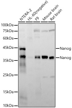
NTERA-2, F9, Mouse brain, Rat brain
Cellular Localization:
Nucleus.
Calculated MW:
35kDa
Observed MW:
37kDa,42kDa
The protein encoded by this gene is a DNA binding homeobox transcription factor involved in embryonic stem (ES) cell proliferation, renewal, and pluripotency. The encoded protein can block ES cell differentiation and can also autorepress its own expression in differentiating cells. Two transcript variants encoding different isoforms have been found for this gene.
Purification Method
Affinity purification
Gene ID
79923
Buffer Information
Store at -20℃. Avoid freeze / thaw cycles. Buffer: PBS with 0.09% Sodium azide,0.05% BSA,50% glycerol,pH7.3.
Western blot analysis of various lysates, using Nanog Rabbit mAb (CAB22625) at 1:10000 dilution. Secondary antibody: HRP-conjugated Goat anti-Rabbit IgG (H+L) (CABS014) at 1:10000 dilution. Lysates/proteins: 25μg per lane. Blocking buffer: 3% nonfat dry milk in TBST. Detection: ECL Enhanced Kit (AbGn00021). Negative control (NC): HL-60. Exposure time: 60s.
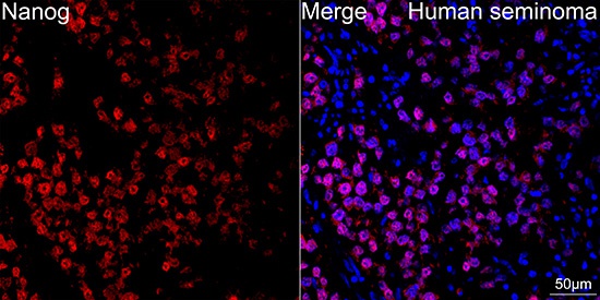
Confocal imaging of paraffin-embedded Human seminoma tissue using Nanog Rabbit mAb (CAB22625, dilution 1:200) followed by a further incubation with Cy3 Goat Anti-Rabbit IgG (H+L) (CABS007, dilution 1:500) (Red). DAPI was used for nuclear staining (Blue). High pressure antigen retrieval performed with 0.01M Citrate Buffer(pH 6.0) prior to IF staining. Objective: 40x.
 at 1:10000 dilution. Secondary antibody: HRP Goat Anti-Rabbit IgG (H+L) at 1:10000 dilution. Lysates/proteins: 25μg per lane. Blocking buffer: 3% nonfat dry milk in TBST.")
 at 1:10000 dilution. Secondary antibody: HRP Goat Anti-Rabbit IgG (H+L) at 1:10000 dilution. Lysates/proteins: 25μg per lane. Blocking buffer: 3% nonfat dry milk in TBST.")
![Anti-Nanog [R01-6K9] Monoclonal Antibody (AGMB00244)](https://cdn11.bigcommerce.com/s-h68l9z2lnx/images/stencil/590x590/products/271533/693214/anti-nanog-r01-6k9-monoclonal-antibody-agmb00244__84172.1774508608.jpg?c=2 "Anti-Nanog [R01-6K9] Monoclonal Antibody (AGMB00244)")
![Anti-Nanog [R05-7S-7] Monoclonal Antibody (AGMB03689)](https://cdn11.bigcommerce.com/s-h68l9z2lnx/images/stencil/590x590/products/274978/680353/anti-nanog-r05-7s-7-monoclonal-antibody-agmb03689__67968.1773041791.jpg?c=2 "Anti-Nanog [R05-7S-7] Monoclonal Antibody (AGMB03689)")

 ELISA Kit (MOEB1300)")
 ELISA Kit (AEKE02366)")