The NHLRC1 Antibody (CAB6669) is a high-quality antibody developed for reliable detection and analysis of target proteins. This polyclonal antibody, produced in rabbits, exhibits high reactivity with human samples and has been validated for use in Western blot applications. By binding to NHLRC1, this antibody enables the detection and analysis of the protein in a variety of cell types, making it an ideal choice for studies in cell biology and neurodegenerative diseases.NHLRC1, also known as EPM2A interacting protein 2, is known to play a key role in the clearance of damaged organelles and proteins through autophagy.
This antibody is validated for use in WB, IHC-P, ELISA applications and has demonstrated reactivity against Human, Mouse, Rat samples.
Product Name:
NHLRC1 Antibody
SKU:
CAB6669
Size:
20μL, 100μL
Reactivity:
Human, Mouse, Rat
Conjugate:
Unconjugated
Immunogen:
Recombinant protein (or fragment).This information is considered to be commercially sensitive.
Recommended starting concentration is 1 μg/mL. Please optimize the concentration based on your specific assay requirements.
Synonyms:
EPM2A, EPM2B, MALIN, bA204B7.2, NHLRC1
Positive Sample:
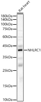
Rat heart
Cellular Localization:
Endoplasmic Reticulum, Nucleus.
Calculated MW:
42kDa
Observed MW:
42kDa/45kDa
The protein encoded by this gene is a single subunit E3 ubiquitin ligase. Laforin is polyubiquitinated by the encoded protein. Defects in this intronless gene lead to an accumulation of laforin and onset of Lafora disease, also known as progressive myoclonic epilepsy type 2 (EPM2).
Purification Method
Affinity purification
Gene ID
378884
RRID
AB_2767255
Buffer Information
Store at -20℃. Avoid freeze / thaw cycles. Buffer: PBS containing 50% glycerol, preserved with proclin300 or sodium azide, pH 7.3.
Western blot analysis of lysates from Rat heart, using NHLRC1 Rabbit pAb (CAB6669) at 1:6000 dilution. Secondary antibody: HRP-conjugated Goat anti-Rabbit IgG (H+L) (CABS014) at 1:10000 dilution. Lysates/proteins: 25μg per lane. Blocking buffer: 3% nonfat dry milk in TBST. Detection: ECL Basic Kit (AbGn00020). Exposure time: 60s.
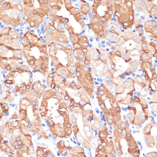
Immunohistochemistry analysis of paraffin-embedded Human stomach using NHLRC1 Rabbit pAb (CAB6669) at dilution of 1:100 (40x lens). Microwave antigen retrieval performed with 0.01M PBS Buffer (pH 7.2) prior to IHC staining.