The p70 S6 Kinase 1 Monoclonal Antibody (CAB4898) is a high-quality antibody developed for reliable detection and analysis of target proteins. This antibody, produced in rabbits, is highly specific to human samples and is validated for use in Western blot applications. By binding to the p70 S6 Kinase protein, this antibody enables accurate detection and analysis in a variety of cell types, making it ideal for studies in cell signaling, cancer research, and drug development.The p70 S6 Kinase protein is a crucial component of the PI3K/Akt/mTOR signaling pathway, which plays a central role in regulating cell proliferation, survival, and metabolism.
This antibody is validated for use in WB, IF/ICC, ELISA applications and has demonstrated reactivity against Human, Mouse, Rat samples.
Product Name:
p70 S6 Kinase 1 Monoclonal Antibody
SKU:
CAB4898
Size:
20μL, 100μL
Reactivity:
Human, Mouse, Rat
Clone Number:
ARC57223
Conjugate:
Unconjugated
Immunogen:
Synthetic peptide. This information is considered to be commercially sensitive.
This gene encodes a member of the ribosomal S6 kinase family of serine/threonine kinases. The encoded protein responds to mTOR (mammalian target of rapamycin) signaling to promote protein synthesis, cell growth, and cell proliferation. Activity of this gene has been associated with human cancer. Alternatively spliced transcript variants have been observed. The use of alternative translation start sites results in isoforms with longer or shorter N-termini which may differ in their subcellular localizations. There are two pseudogenes for this gene on chromosome 17.
Purification Method
Affinity purification
Gene ID
6198
RRID
AB_2863381
Buffer Information
Store at -20℃. Avoid freeze / thaw cycles. Buffer: PBS containing 50% glycerol and 0.05% BSA, preserved with proclin300 or sodium azide, pH 7.3.
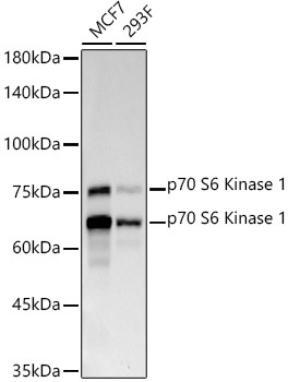
Western blot analysis of various lysates using p70 S6 Kinase 1 Rabbit mAb (CAB4898) at 1:1000 dilution incubated overnight at 4℃. Secondary antibody: HRP-conjugated Goat anti-Rabbit IgG (H+L) (CABS014) at 1:10000 dilution. Lysates/proteins: 25μg per lane. Blocking buffer: 3% nonfat dry milk in TBST. Detection: ECL Basic Kit (AbGn00020). Exposure time: 30s.
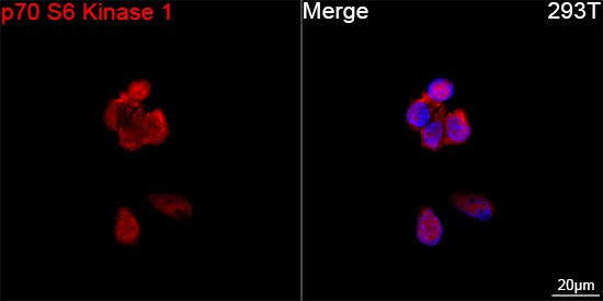
Confocal imaging of 293T cells using p70 S6 Kinase 1 Rabbit mAb (CAB4898, dilution 1:200) followed by a further incubation with Cy3 Goat Anti-Rabbit IgG (H+L) (CABS007, dilution 1:500) (Red). DAPI was used for nuclear staining (Blue). Objective: 100x.
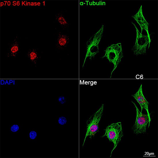
Confocal imaging of C6 cells using p70 S6 Kinase 1 Rabbit mAb (CAB4898, dilution 1:200) followed by a further incubation with Cy3 Goat Anti-Rabbit IgG (H+L) (CABS007, dilution 1:500) (Red). The cells were counterstained with α-Tubulin Mouse mAb (AC012, dilution 1:400) followed by incubation with ABflo® 488-conjugated Goat Anti-Mouse IgG (H+L) Ab (CABS076, dilution 1:500) (Green). DAPI was used for nuclear staining (Blue). Objective: 100x.