The PAK1 Monoclonal Antibody (CAB19608) is a high-quality antibody developed for reliable detection and analysis of target proteins. This antibody, generated from rabbit cells, exhibits high reactivity towards human samples and has been validated for use in Western blot applications. By specifically binding to the PAK1 protein, this antibody enables accurate detection and analysis in a variety of cell types, making it ideal for studies in cancer biology, cell biology, and signal transduction pathways.PAK1, a member of the p21-activated kinase (PAK) family, plays a crucial role in regulating cell growth, division, and movement.
This antibody is validated for use in WB, IF/ICC, ELISA applications and has demonstrated reactivity against Human, Mouse, Rat samples.
Product Name:
PAK1 Monoclonal Antibody
SKU:
CAB19608
Size:
20μL, 100μL
Reactivity:
Human, Mouse, Rat
Clone Number:
ARC0087
Conjugate:
Unconjugated
Immunogen:
Synthetic peptide. This information is considered to be commercially sensitive.
This gene encodes a family member of serine/threonine p21-activating kinases, known as PAK proteins. These proteins are critical effectors that link RhoGTPases to cytoskeleton reorganization and nuclear signaling, and they serve as targets for the small GTP binding proteins Cdc42 and Rac. This specific family member regulates cell motility and morphology. Mutations in this gene have been associated with macrocephaly, seizures, and speech delay. Overexpression of this gene is also reported in many cancer types, and particularly in breast cancer. Alternative splicing results in multiple transcript variants.
Purification Method
Affinity purification
Gene ID
5058
RRID
AB_2862697
Buffer Information
Store at -20℃. Avoid freeze / thaw cycles. Buffer: PBS containing 50% glycerol and 0.05% BSA, preserved with proclin300 or sodium azide, pH 7.3.
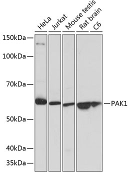
Western blot analysis of various lysates using PAK1 Rabbit mAb (CAB19608) at 1:1000 dilution. Secondary antibody: HRP-conjugated Goat anti-Rabbit IgG (H+L) (CABS014) at 1:10000 dilution. Lysates/proteins: 25μg per lane. Blocking buffer: 3% nonfat dry milk in TBST. Detection: ECL Basic Kit (AbGn00020). Exposure time: 1s.
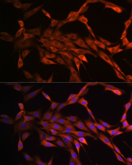
Immunofluorescence analysis of NIH/3T3 cells using PAK1 Rabbit mAb (CAB19608) at dilution of 1:100 (40x lens). Secondary antibody: Cy3-conjugated Goat anti-Rabbit IgG (H+L) (CABS007) at 1:500 dilution. Blue: DAPI for nuclear staining.