The Pan-AKT Antibody (CAB18120) is a high-quality antibody developed for reliable detection and analysis of target proteins. This rabbit polyclonal antibody is highly specific and reactive with human samples, making it ideal for use in Western blot applications to detect and analyze AKT protein levels.AKT, also known as protein kinase B, plays a critical role in various cellular processes, including cell cycle progression, apoptosis, and metabolism. Dysregulation of the AKT pathway has been implicated in various diseases, including cancer, diabetes, and neurological disorders.
This antibody is validated for use in WB, IHC-P, IF/ICC, IP, ELISA applications and has demonstrated reactivity against Human, Mouse, Rat samples.
Product Name:
Pan-AKT Antibody
SKU:
CAB18120
Size:
20μL, 100μL
Reactivity:
Human, Mouse, Rat
Conjugate:
Unconjugated
Immunogen:
Recombinant protein (or fragment).This information is considered to be commercially sensitive.
Human AKT serine-threonine protein kinase family includes three members AKT1,AKT2, AKT3, which are also often referred to as protein kinase B alpha, beta, and gamma. These highly similar AKT proteins all have an N-terminal pleckstrin homology domain, a serine/threonine-specific kinase domain and a C-terminal regulatory domain. These proteins are phosphorylated by phosphoinositide 3-kinase (PI3K). AKT/PI3K forms a key component of many signalling pathways that involve the binding of membrane-bound ligands such as receptor tyrosine kinases, G-protein coupled receptors, and integrin-linked kinase. These AKT proteins therefore regulate a wide variety of cellular functions including cell proliferation, survival, metabolism, and angiogenesis in both normal and malignant cells. AKT proteins are recruited to the cell membrane by phosphatidylinositol 3,4,5-trisphosphate (PIP3) after phosphorylation of phosphatidylinositol 4,5-bisphosphate (PIP2) by PI3K. Subsequent phosphorylation of both threonine residue 308 and serine residue 473 is required for full activation of the AKT1 protein encoded by this gene.
Purification Method
Affinity purification
Gene ID
207 208 10000
RRID
AB_2861912
Buffer Information
Store at -20℃. Avoid freeze / thaw cycles. Buffer: PBS containing 50% glycerol, preserved with proclin300 or sodium azide, pH 7.3.
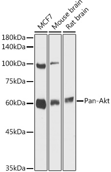
Western blot analysis of various lysates using Pan-Akt Rabbit pAb (CAB18120) at 1:1000 dilution. Secondary antibody: HRP-conjugated Goat anti-Rabbit IgG (H+L) (CABS014) at 1:10000 dilution. Lysates/proteins: 25μg per lane. Blocking buffer: 3% nonfat dry milk in TBST. Detection: ECL Basic Kit (AbGn00020). Exposure time: 30s.
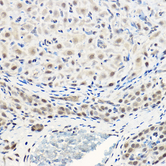
Immunohistochemistry analysis of paraffin-embedded Rat ovary using Pan-Akt Rabbit pAb (CAB18120) at dilution of 1:100 (40x lens). High pressure antigen retrieval performed with 0.01M Citrate buffer (pH 6.0) prior to IHC staining.
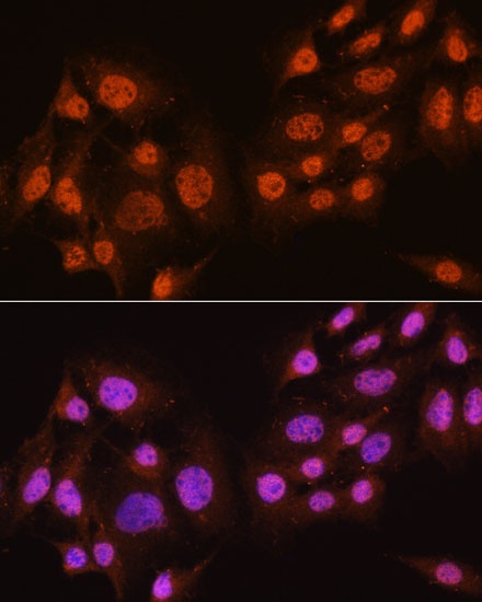
Immunofluorescence analysis of C6 cells using Pan-Akt Rabbit pAb (CAB18120) at dilution of 1:100. Secondary antibody: Cy3-conjugated Goat anti-Rabbit IgG (H+L) (CABS007) at 1:500 dilution. Blue: DAPI for nuclear staining.
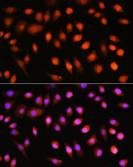
Immunofluorescence analysis of L929 cells using Pan-Akt Rabbit pAb (CAB18120) at dilution of 1:100. Secondary antibody: Cy3-conjugated Goat anti-Rabbit IgG (H+L) (CABS007) at 1:500 dilution. Blue: DAPI for nuclear staining.
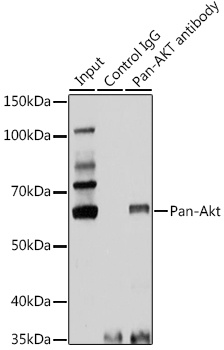
Immunoprecipitation analysis of 25 μg extracts of Rat brain cells using 3 μg Pan-Akt antibody (CAB18120). Western blot was performed from the immunoprecipitate using Pan-Akt antibody (CAB18120) at a dilution of 1:1000.