The PDXK Polyclonal Antibody (CAB24034) is a high-quality antibody developed for reliable detection and analysis of target proteins. This antibody, generated in rabbits, is highly specific to human samples and is validated for use in Western blot applications. By binding to the PDXK protein, this antibody enables the detection and analysis of PDXK in various cell types, making it ideal for studies in biochemistry, metabolism, and cancer research.PDXK is a key player in vitamin B6 metabolism, converting pyridoxine (vitamin B6) into its active form, pyridoxal phosphate. This process is crucial for a variety of cellular functions, including amino acid metabolism, neurotransmitter synthesis, and DNA synthesis.
This antibody is validated for use in WB, IF/ICC, ELISA applications and has demonstrated reactivity against Human, Mouse, Rat samples.
Product Name:
PDXK Polyclonal Antibody
SKU:
CAB24034
Size:
20μL, 100μL
Reactivity:
Human, Mouse, Rat
Conjugate:
Unconjugated
Immunogen:
Synthetic peptide. This information is considered to be commercially sensitive.
NIH/3T3, Mouse kidney, Rat liver, HeLa, RT4, NIH/3T3, Mouse kidney, Rat kidney
Cellular Localization:
Cytoplasm.
Calculated MW:
35kDa
Observed MW:
35kDa
The protein encoded by this gene phosphorylates vitamin B6, a step required for the conversion of vitamin B6 to pyridoxal-5-phosphate, an important cofactor in intermediary metabolism. The encoded protein is cytoplasmic and probably acts as a homodimer. Alternatively spliced transcript variants have been described, but their biological validity has not been determined.
Purification Method
Affinity purification
Gene ID
8566
Buffer Information
Store at -20℃. Avoid freeze / thaw cycles. Buffer: PBS containing 50% glycerol, preserved with proclin300 or sodium azide, pH 7.3.
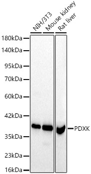
Western blot analysis of various lysates using [KD Validated]PDXK Rabbit pAb (CAB24034) at 1:1000 dilution. Secondary antibody: HRP-conjugated Goat anti-Rabbit IgG (H+L) (CABS014) at 1:10000 dilution. Lysates / proteins: 25 μg per lane. Blocking buffer: 3 % nonfat dry milk in TBST. Detection: ECL Basic Kit (AbGn00020). Exposure time: 30s.
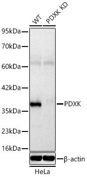
Western blot analysis of lysates from wild type (WT) and PDXK knockdown (KD) HeLa cells using [KD Validated]PDXK Rabbit pAb (CAB24034) at 1:1600 dilution incubated overnight at 4℃. Secondary antibody: HRP-conjugated Goat anti-Rabbit IgG (H+L) (CABS014) at 1:10000 dilution. Lysates/proteins: 25 μg per lane. Blocking buffer: 3% nonfat dry milk in TBST. Detection: ECL Basic Kit (AbGn00020). Exposure time: 10s.
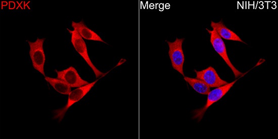
Immunofluorescence analysis of NIH/3T3 cells using [KD Validated]PDXK Rabbit pAb (CAB24034) at a dilution of 1:100 (40x lens). Secondary antibody: Cy3-conjugated Goat anti-Rabbit IgG (H+L) (CABS007) at 1:500 dilution. Blue: DAPI for nuclear staining.
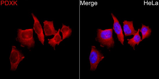
Immunofluorescence analysis of HeLa cells using [KD Validated]PDXK Rabbit pAb (CAB24034) at a dilution of 1:100 (40x lens). Secondary antibody: Cy3-conjugated Goat anti-Rabbit IgG (H+L) (CABS007) at 1:500 dilution. Blue: DAPI for nuclear staining.
")
")


![Anti- PDXK [5H5] Monoclonal Antibody - Knockout Validated (AGMB06760)](https://cdn11.bigcommerce.com/s-h68l9z2lnx/images/stencil/590x590/products/278041/731477/anti-pdxk-5h5-monoclonal-antibody-knockout-validated-agmb06760__48115.1777183779.jpg?c=2 "Anti- PDXK [5H5] Monoclonal Antibody - Knockout Validated (AGMB06760)")

")