The PHC1 Antibody (CAB5843) is a high-quality antibody developed for reliable detection and analysis of target proteins. This antibody, developed through rabbit immunization, exhibits high reactivity with human samples and is validated for use in Western blotting applications.PHC1, a component of the polycomb repressive complex 1 (PRC1), is involved in the epigenetic control of gene expression through chromatin remodeling.
This antibody is validated for use in WB, ELISA applications and has demonstrated reactivity against Human, Mouse, Rat samples.
Product Name:
PHC1 Antibody
SKU:
CAB5843
Size:
20μL, 100μL
Reactivity:
Human, Mouse, Rat
Conjugate:
Unconjugated
Immunogen:
Recombinant protein (or fragment).This information is considered to be commercially sensitive.
Recommended starting concentration is 1 μg/mL. Please optimize the concentration based on your specific assay requirements.
Synonyms:
EDR1, HPH1, RAE28, MCPH11, PHC1
Positive Sample:
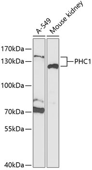
A-549, Mouse kidney
Cellular Localization:
Nucleus.
Calculated MW:
106kDa
Observed MW:
125kDa/135kDa
This gene is a homolog of the Drosophila polyhomeotic gene, which is a member of the Polycomb group of genes. The gene product is a component of a multimeric protein complex that contains EDR2 and the vertebrate Polycomb protein BMH1. The gene product, the EDR2 protein, and the Drosophila polyhomeotic protein share 2 highly conserved domains, named homology domains I and II. These domains are involved in protein-protein interactions and may mediate heterodimerization of the protein encoded by this gene and the EDR2 protein.
Purification Method
Affinity purification
Gene ID
1911
RRID
AB_2766593
Buffer Information
Store at -20℃. Avoid freeze / thaw cycles. Buffer: PBS containing 50% glycerol, preserved with proclin300 or sodium azide, pH 7.3.
Western blot analysis of various lysates using PHC1 Rabbit pAb (CAB5843) at 1:1000 dilution. Secondary antibody: HRP-conjugated Goat anti-Rabbit IgG (H+L) (CABS014) at 1:10000 dilution. Lysates/proteins: 25μg per lane. Blocking buffer: 3% nonfat dry milk in TBST. Detection: ECL Enhanced Kit (AbGn00021). Exposure time: 90s.