The PHIP Antibody (CAB7207) is a high-quality antibody developed for reliable detection and analysis of target proteins. This antibody, produced in rabbits, exhibits high reactivity with human samples and is validated for use in Western blot applications. By targeting the PHIP protein, this antibody enables precise detection and analysis in a variety of cell types, making it an ideal choice for investigations in the fields of metabolism, obesity, and diabetes research.PHIP, also known as pleckstrin homology domain-interacting protein, is involved in regulating insulin signaling and glucose metabolism, making it a key player in metabolic disorders and related conditions.
This antibody is validated for use in WB, IHC-P, IF/ICC, ELISA applications and has demonstrated reactivity against Human, Mouse samples.
Product Name:
PHIP Antibody
SKU:
CAB7207
Size:
20μL, 100μL
Reactivity:
Human, Mouse
Conjugate:
Unconjugated
Immunogen:
Recombinant protein (or fragment).This information is considered to be commercially sensitive.
Recommended starting concentration is 1 μg/mL. Please optimize the concentration based on your specific assay requirements.
Synonyms:
ndrp, BRWD2, DIDOD, WDR11, DCAF14, CHUJANS, PHIP
Positive Sample:
HeLa, Jurkat
Cellular Localization:
Nucleus.
Calculated MW:
207kDa
Observed MW:
250kDa
This gene encodes a protein that binds to the insulin receptor substrate 1 protein and regulates glucose transporter translocation in skeletal muscle cells. The encoded protein may also regulate growth and survival of pancreatic beta cells. Elevated copy number of this gene may be associated with melanoma severity and the encoded protein may promote melanoma metastasis in human patients.
Purification Method
Affinity purification
Gene ID
55023
RRID
AB_2767756
Buffer Information
Store at -20℃. Avoid freeze / thaw cycles. Buffer: PBS containing 50% glycerol, preserved with proclin300 or sodium azide, pH 7.3.
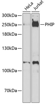
Western blot analysis of various lysates using PHIP Rabbit pAb (CAB7207) at 1:1000 dilution._Secondary antibody: HRP-conjugated Goat anti-Rabbit IgG (H+L) (CABS014) at 1:10000 dilution._Lysates/proteins: 25μg per lane._Blocking buffer: 3% nonfat dry milk in TBST._Detection: ECL Enhanced Kit (AbGn00021)._Exposure time: 90s.
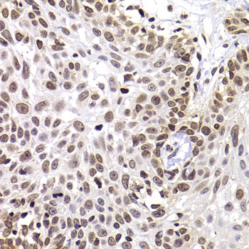
Immunohistochemistry analysis of paraffin-embedded Human well-differentiated squamous skin carcinoma using PHIP Rabbit pAb (CAB7207) at dilution of 1:100 (40x lens). Microwave antigen retrieval performed with 0.01M PBS Buffer (pH 7.2) prior to IHC staining.
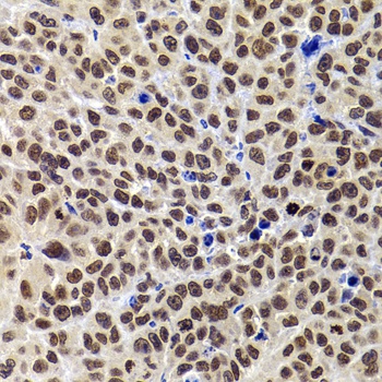
Immunohistochemistry analysis of paraffin-embedded Mouse tumor using PHIP Rabbit pAb (CAB7207) at dilution of 1:100 (40x lens). Microwave antigen retrieval performed with 0.01M PBS Buffer (pH 7.2) prior to IHC staining.
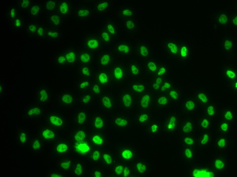
Immunofluorescence analysis of MCF-7 cells using PHIP Rabbit pAb (CAB7207).Secondary antibody: Cy3-conjugated Goat anti-Rabbit IgG (H+L) (CABS007) at 1:500 dilution.