The PHLDA2 Antibody (CAB6244) is a high-quality antibody developed for reliable detection and analysis of target proteins. This antibody, produced in rabbits, is highly specific for human samples and has been validated for use in Western blot applications. By binding to the PHLDA2 protein, this antibody enables researchers to detect and analyze PHLDA2 expression in various cell types, making it an essential component for studies in cancer biology and cell signaling pathways.PHLDA2, also known as T-cell death-associated gene 51 (TDAG51), is a crucial regulator of cell growth and survival, with implications in cancer development and progression.
This antibody is validated for use in WB, IF/ICC, ELISA applications and has demonstrated reactivity against Human, Mouse samples.
Product Name:
PHLDA2 Antibody
SKU:
CAB6244
Size:
20μL, 100μL
Reactivity:
Human, Mouse
Conjugate:
Unconjugated
Immunogen:
Recombinant protein (or fragment).This information is considered to be commercially sensitive.
Recommended starting concentration is 1 μg/mL. Please optimize the concentration based on your specific assay requirements.
Synonyms:
IPL, BRW1C, BWR1C, HLDA2, TSSC3, PHLDA2
Positive Sample:
PC-3
Cellular Localization:
Cytoplasm, Membrane, Peripheral Membrane Protein.
Calculated MW:
17kDa
Observed MW:
20kDa
This gene is located in a cluster of imprinted genes on chromosome 11p15.5, which is considered to be an important tumor suppressor gene region. Alterations in this region may be associated with the Beckwith-Wiedemann syndrome, Wilms tumor, rhabdomyosarcoma, adrenocortical carcinoma, and lung, ovarian, and breast cancer. This gene has been shown to be imprinted, with preferential expression from the maternal allele in placenta and liver.
Purification Method
Affinity purification
Gene ID
7262
RRID
AB_2766851
Buffer Information
Store at -20℃. Avoid freeze / thaw cycles. Buffer: PBS containing 50% glycerol, preserved with proclin300 or sodium azide, pH 7.3.
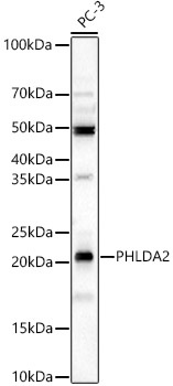
Western blot analysis of lysates from PC-3 cells using PHLDA2 Rabbit pAb (CAB6244) at 1:4000 dilution. Secondary antibody: HRP-conjugated Goat anti-Rabbit IgG (H+L) (CABS014) at 1:10000 dilution. Lysates/proteins: 25 μg per lane. Blocking buffer: 3% nonfat dry milk in TBST. Detection: ECL Basic Kit (AbGn00020). Exposure time:180s.
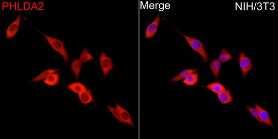
Immunofluorescence analysis of NIH/3T3 cells using PHLDA2 Rabbit pAb (CAB6244) at dilution of 1:500 (40x lens). Secondary antibody: Cy3-conjugated Goat anti-Rabbit IgG (H+L) (CABS007) at 1:500 dilution. Blue: DAPI for nuclear staining.