The Phospho-ATR-S428 Antibody (CABP0676) is a high-quality antibody developed for reliable detection and analysis of target proteins. This antibody, produced in rabbits, is highly specific for detecting phosphorylated ATR at serine 428 in human samples. It has been validated for use in Western blot applications, enabling researchers to assess ATR activation in various cell types.The phosphorylation of ATR at serine 428 is known to play a crucial role in the activation of the ATR signaling pathway in response to DNA damage. By targeting this specific phosphorylation site, researchers can gain insights into the mechanisms underlying DNA damage repair and cell cycle checkpoint control.
This antibody is validated for use in WB, IHC-P, IF/ICC, ELISA applications and has demonstrated reactivity against Human, Mouse, Rat samples.
Product Name:
Phospho-ATR-S428 Antibody
SKU:
CABP0676
Size:
20μL, 100μL
Reactivity:
Human, Mouse, Rat
Conjugate:
Unconjugated
Immunogen:
Synthetic peptide. This information is considered to be commercially sensitive.
Sequence:
DGIS PK
Tested Applications:
WBIHC-PIF/ICCELISA
Recommended Dilution:
WB
1:100 - 1:500
IHC-P
1:50 - 1:200
IF/ICC
1:50 - 1:200
ELISA
Recommended starting concentration is 1 μg/mL. Please optimize the concentration based on your specific assay requirements.
Synonyms:
FRP1, MEC1, SCKL, FCTCS, SCKL1, Phospho-ATR-S428
Positive Sample:
293T treated with UV, NIH/3T3 treated with Nocodazole
Cellular Localization:
Chromosome, Nucleus, Pml Body.
Calculated MW:
301kDa
Observed MW:
300kDa
The protein encoded by this gene is a serine/threonine kinase and DNA damage sensor, activating cell cycle checkpoint signaling upon DNA stress. The encoded protein can phosphorylate and activate several proteins involved in the inhibition of DNA replication and mitosis, and can promote DNA repair, recombination, and apoptosis. This protein is also important for fragile site stability and centrosome duplication. Defects in this gene are a cause of Seckel syndrome 1.
Purification Method
Affinity purification
Gene ID
545
RRID
AB_2770928
Buffer Information
Store at -20℃. Avoid freeze / thaw cycles. Buffer: PBS containing 50% glycerol, preserved with proclin300 or sodium azide, pH 7.3.
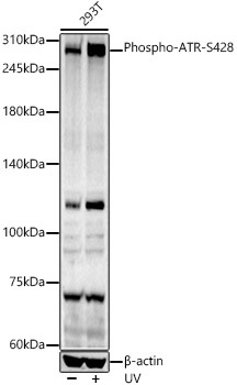
Western blot analysis of lysates from 293T cells, using Phospho-ATR-S428 Rabbit pAb (CABP0676) at 1:400 dilution. 293T cells were treated with UV at room temperature for 15-30 minutes. Secondary antibody: HRP-conjugated Goat anti-Rabbit IgG (H+L) (CABS014) at 1:10000 dilution. Lysates/proteins: 25μg per lane. Blocking buffer: 3% nonfat dry milk in TBST. Detection: ECL Basic Kit (AbGn00020). Exposure time: 90s.
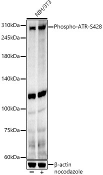
Western blot analysis of lysates from NIH/3T3 cells, using Phospho-ATR-S428 Rabbit pAb (CABP0676) at 1:400 dilution. NIH/3T3 cells were treated with Nocodazole (50 ng/ml) at 37℃ for 20 hours. Secondary antibody: HRP-conjugated Goat anti-Rabbit IgG (H+L) (CABS014) at 1:10000 dilution. Lysates/proteins: 25μg per lane. Blocking buffer: 3% nonfat dry milk in TBST. Detection: ECL Basic Kit (AbGn00020). Exposure time: 90s.
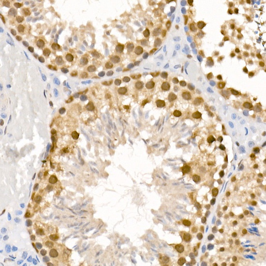
Immunohistochemistry analysis of paraffin-embedded Mouse testis using Phospho-ATR-S428 Rabbit pAb (CABP0676) at dilution of 1:100 (40x lens). High pressure antigen retrieval performed with 0.01M Citrate buffer (pH 6.0) prior to IHC staining.
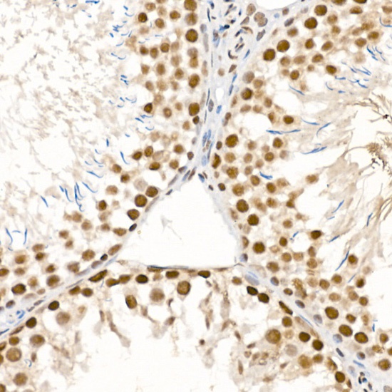
Immunohistochemistry analysis of paraffin-embedded Rat testis using Phospho-ATR-S428 Rabbit pAb (CABP0676) at dilution of 1:100 (40x lens). High pressure antigen retrieval performed with 0.01M Citrate buffer (pH 6.0) prior to IHC staining.