The Phospho-BRD4-T204 Antibody (CABP1124) is a high-quality antibody developed for reliable detection and analysis of target proteins. BRD4 is a key player in epigenetic regulation and gene expression, particularly in cancer cells where it plays a role in cell proliferation and survival.This antibody, raised in rabbits, specifically recognizes the phosphorylated form of BRD4 at threonine 204 in human samples, making it an ideal choice for Western blot applications. By targeting this specific phosphorylation site, researchers can gain insight into the role of BRD4 in cancer progression and potentially identify new therapeutic targets.
This antibody is validated for use in WB, ELISA applications and has demonstrated reactivity against Human, Mouse, Rat samples.
Product Name:
Phospho-BRD4-T204 Antibody
SKU:
CABP1124
Size:
20μL, 100μL
Reactivity:
Human, Mouse, Rat
Immunogen:
Synthetic peptide. This information is considered to be commercially sensitive.
Sequence:
Email for sequence
Tested Applications:
WBELISA
Recommended Dilution:
WB
1:500 - 1:1000
ELISA
Recommended starting concentration is 1 μg/mL. Please optimize the concentration based on your specific assay requirements.
The protein encoded by this gene is homologous to the murine protein MCAP, which associates with chromosomes during mitosis, and to the human RING3 protein, a serine/threonine kinase. Each of these proteins contains two bromodomains, a conserved sequence motif which may be involved in chromatin targeting. This gene has been implicated as the chromosome 19 target of translocation t(15;19)(q13;p13.1), which defines an upper respiratory tract carcinoma in young people. Two alternatively spliced transcript variants have been described.
Purification Method
Affinity purification
Gene ID
23476
RRID
AB_2863992
Buffer Information
Store at -20℃. Avoid freeze / thaw cycles. Buffer: PBS with 0.09% Sodium azide,50% glycerol,pH7.3.
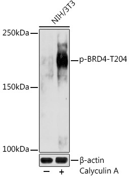
Western blot analysis of lysates from NIH/3T3 cells, using Phospho-BRD4-T204 Rabbit pAb (CABP1124) at 1:1000 dilution. NIH/3T3 cells were treated with Calyculin A (100 nM) at 37℃ for 30 minutes after serum-starvation overnight. Secondary antibody: HRP-conjugated Goat anti-Rabbit IgG (H+L) (CABS014) at 1:10000 dilution. Lysates/proteins: 25μg per lane. Blocking buffer: 3% nonfat dry milk in TBST. Detection: ECL Basic Kit (AbGn00020). Exposure time: 180s.


 for 30 minutes with NaCl (400mM) after serum-starvation 16-20 hours. Secondary antibody: HRP Goat Anti-Rabbit IgG (H+L) at 1:10000 dilution. Lysates/proteins: 25ug per lane. Blocking buffer: 3% nonfat dry milk in TBST. Detection: ECL Enhanced Kit. Exposure time: 90s.")
 at 37℃ for 30 minutes after serum-starvation overnight. C6 cells were treated by Calyculin A (100 nM) at 37℃ for 30 minutes after serum-starvation overnight. Secondary antibody: HRP Goat Anti-Rabbit IgG (H+L) at 1:10000 dilution. Lysates/proteins: 25ug per lane. Blocking buffer: 3% nonfat dry milk in TBST. Detection: ECL Basic Kit. Exposure time: 1s.")
")

![Anti-Brd4 [R01-1H8] Monoclonal Antibody (AGMB04160)](https://cdn11.bigcommerce.com/s-h68l9z2lnx/images/stencil/590x590/products/275449/679793/anti-brd4-r01-1h8-monoclonal-antibody-agmb04160__32522.1773040000.jpg?c=2 "Anti-Brd4 [R01-1H8] Monoclonal Antibody (AGMB04160)")
