The Phospho-eIF4EBP1-S65 Monoclonal Antibody (CABP1363) is a high-quality antibody developed for reliable detection and analysis of target proteins. This antibody, derived from mouse cells, specifically targets the phosphorylated form of EIF4EBP1 at serine 65, allowing for precise detection and analysis in various experimental settings.Phosphorylation of EIF4EBP1 at serine 65 is known to play a crucial role in the regulation of protein translation, making this antibody essential for studying cellular processes such as cell growth, proliferation, and survival.
This antibody is validated for use in WB, ELISA applications and has demonstrated reactivity against Human, Mouse samples.
Product Name:
Phospho-eIF4EBP1-S65 Monoclonal Antibody
SKU:
CABP1363
Size:
20μL, 100μL
Reactivity:
Human, Mouse
Clone Number:
ARC53775
Conjugate:
Unconjugated
Immunogen:
Synthetic peptide. This information is considered to be commercially sensitive.
Sequence:
RNSP V
Tested Applications:
WBELISA
Recommended Dilution:
WB
1:1000 - 1:12000
ELISA
Recommended starting concentration is 1 μg/mL. Please optimize the concentration based on your specific assay requirements.
Synonyms:
BP-1, 4EBP1, 4E-BP1, PHAS-I, Phospho-eIF4EBP1-S65
Positive Sample:
NIH/3T3 treated with λ-pp, 293T treated with insulin
Cellular Localization:
Cytoplasm, Cytosol.
Calculated MW:
13kDa
Observed MW:
15-20kDa
This gene encodes one member of a family of translation repressor proteins. The protein directly interacts with eukaryotic translation initiation factor 4E (eIF4E), which is a limiting component of the multisubunit complex that recruits 40S ribosomal subunits to the 5' end of mRNAs. Interaction of this protein with eIF4E inhibits complex assembly and represses translation. This protein is phosphorylated in response to various signals including UV irradiation and insulin signaling, resulting in its dissociation from eIF4E and activation of mRNA translation.
Purification Method
Affinity purification
Gene ID
1978
Buffer Information
Store at -20℃. Avoid freeze / thaw cycles. Buffer: PBS containing 50% glycerol and 0.05% BSA, preserved with proclin300 or sodium azide, pH 7.3.
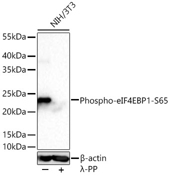
Western blot analysis of lysates from NIH/3T3 cells using Phospho-eIF4EBP1-S65 Rabbit mAb (CABP1363) at 1:1000 dilution incubated at room temperature for 1.5 hours. NIH/3T3 cells were treated with λ-PP mixed solution (1ul) at 30℃ for 30 minutes. Secondary antibody: HRP-conjugated Goat anti-Rabbit IgG (H+L) (CABS014) at 1:10000 dilution. Lysates/proteins: 30 μg per lane. Blocking buffer: 3 % nonfat dry milk in TBST. Detection: ECL Basic Kit (AbGn00020). Exposure time: 90s.
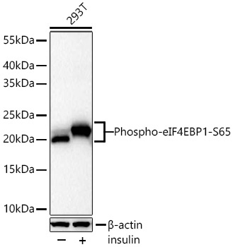
Western blot analysis of lysates from 293T cells using Phospho-eIF4EBP1-S65 Rabbit mAb (CABP1363) at 1:12000 dilution incubated at room temperature for 1.5 hours. 293T cells were treated with insulin (100 ng/ml) at 37℃ for 40 minutes after serum-starvation overnight. Secondary antibody: HRP-conjugated Goat anti-Rabbit IgG (H+L) (CABS014) at 1:10000 dilution. Lysates/proteins: 30 μg per lane. Blocking buffer: 3 % nonfat dry milk in TBST. Detection: ECL Basic Kit (AbGn00020). Exposure time: 90s.
 at 1:20000 dilution. 293T cells were treated by IGF-1 (50 ng/ml) at 37℃ for 5 minutes after serum-starvation overnight. Secondary antibody: HRP Goat Anti-Rabbit IgG (H+L) at 1:10000 dilution. Lysates/proteins: 25μg per lane. Blocking buffer: 3% nonfat dry milk in TBST.")
 at 1:20000 dilution. 293T cells were treated by IGF-1 (50 ng/ml) at 37℃ for 5 minutes after serum-starvation overnight. Secondary antibody: HRP Goat Anti-Rabbit IgG (H+L) at 1:10000 dilution. Lysates/proteins: 25μg per lane. Blocking buffer: 3% nonfat dry milk in TBST.")