The Phospho-HSP27/HSPB1-S82 Antibody (CABP0041) is a high-quality antibody developed for reliable detection and analysis of target proteins. This antibody, generated in rabbits, is highly specific to the phosphorylated form of HSPB1 and is suitable for use in Western blot applications with human samples.HSPB1 is a chaperone protein that plays a key role in cellular stress response and is involved in various cellular processes, including cell survival, apoptosis, and cytoskeletal organization. Phosphorylation of HSPB1 at serine 82 has been linked to its function in regulating actin dynamics and cytoskeletal rearrangements in response to stress stimuli.
This antibody is validated for use in WB, IHC-P, IP, ELISA applications and has demonstrated reactivity against Human samples.
Product Name:
Phospho-HSP27/HSPB1-S82 Antibody
SKU:
CABP0041
Size:
20μL, 100μL
Reactivity:
Human
Conjugate:
Unconjugated
Immunogen:
Synthetic peptide. This information is considered to be commercially sensitive.
Sequence:
QLSS G
Tested Applications:
WBIHC-PIPELISA
Recommended Dilution:
WB
1:500 - 1:2000
IHC-P
1:50 - 1:200
IP
0.5μg-4μg antibody for 200μg-400μg extracts of whole cells
ELISA
Recommended starting concentration is 1 μg/mL. Please optimize the concentration based on your specific assay requirements.
HeLa treated with EGF, HeLa treated with UV, HeLa treated with Anisomycin
Cellular Localization:
Cytoplasm, Nucleus, Cytoskeleton, Spindle.
Calculated MW:
23kDa
Observed MW:
28kDa
This gene encodes a member of the small heat shock protein (HSP20) family of proteins. In response to environmental stress, the encoded protein translocates from the cytoplasm to the nucleus and functions as a molecular chaperone that promotes the correct folding of other proteins. This protein plays an important role in the differentiation of a wide variety of cell types. Expression of this gene is correlated with poor clinical outcome in multiple human cancers, and the encoded protein may promote cancer cell proliferation and metastasis, while protecting cancer cells from apoptosis. Mutations in this gene have been identified in human patients with Charcot-Marie-Tooth disease and distal hereditary motor neuropathy.
Purification Method
Affinity purification
Gene ID
3315
RRID
AB_2771186
Buffer Information
Store at -20℃. Avoid freeze / thaw cycles. Buffer: PBS with 0.09% Sodium azide,50% glycerol,pH7.3.
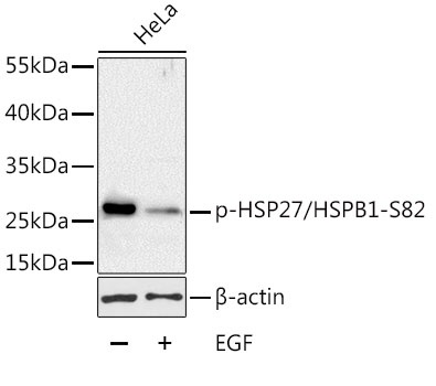
Western blot analysis of lysates from HeLa cells, using Phospho-HSP27/HSPB1-S82 Rabbit pAb (CABP0041) at 1:1000 dilution. HeLa cells were treated with EGF (100ng/mL) for 30 minutes after serum-starvation overnight. Secondary antibody: HRP-conjugated Goat anti-Rabbit IgG (H+L) (CABS014) at 1:10000 dilution. Lysates/proteins: 25μg per lane. Blocking buffer: 3% BSA. Detection: ECL Basic Kit (AbGn00020). Exposure time: 1min.
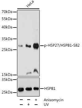
Western blot analysis of lysates from HeLa cells, using Phospho-HSP27/HSPB1-S82 pAb (CABP0041) at 1:1000 dilution or HSP27/HSPB1 antibody (CAB16332). HeLa cells were treated with UV at room temperature for 15-30 minutes.HeLa cells were treated with Anisomycin (25 μg/mL) at 37℃ for 30 minutes after serum-starvation overnight. Secondary antibody: HRP-conjugated Goat anti-Rabbit IgG (H+L) (CABS014) at 1:10000 dilution. Lysates/proteins: 25μg per lane. Blocking buffer: 3% BSA. Detection: ECL Basic Kit (AbGn00020). Exposure time: 10s.