The Phospho-POLR2A-S5 Monoclonal Antibody (CABP0997) is a high-quality antibody developed for reliable detection and analysis of target proteins. This antibody, generated in rabbits, exhibits high specificity and sensitivity for detecting phosphorylated POLR2A in human samples, making it an excellent choice for Western blot applications.Phosphorylation of POLR2A at serine 5 is a critical step in the transcription process, influencing RNA polymerase II activity and gene expression. By targeting this specific phosphorylation site, researchers can gain valuable insights into the molecular mechanisms underlying gene regulation and transcriptional control.
This antibody is validated for use in WB, IHC-P, IF/ICC, ELISA applications and has demonstrated reactivity against Human, Mouse, Rat samples.
Product Name:
Phospho-POLR2A-S5 Monoclonal Antibody
SKU:
CABP0997
Size:
20μL, 100μL
Reactivity:
Human, Mouse, Rat
Clone Number:
ARC1541
Conjugate:
Unconjugated
Immunogen:
Synthetic peptide. This information is considered to be commercially sensitive.
Tested Applications:
WBIHC-PIF/ICCELISA
Recommended Dilution:
WB
1:1000 - 1:4000
IHC-P
1:50 - 1:200
IF/ICC
1:50 - 1:200
ELISA
Recommended starting concentration is 1 μg/mL. Please optimize the concentration based on your specific assay requirements.
MCF7 treated with Doxorubicin, MCF7 treated with nocodazole, C2C12
Cellular Localization:
Nucleus.
Calculated MW:
217kDa
Observed MW:
270kDa
This gene encodes the largest subunit of RNA polymerase II, the polymerase responsible for synthesizing messenger RNA in eukaryotes. The product of this gene contains a carboxy terminal domain composed of heptapeptide repeats that are essential for polymerase activity. These repeats contain serine and threonine residues that are phosphorylated in actively transcribing RNA polymerase. In addition, this subunit, in combination with several other polymerase subunits, forms the DNA binding domain of the polymerase, a groove in which the DNA template is transcribed into RNA.
Purification Method
Affinity purification
Gene ID
5430
RRID
AB_2863889
Buffer Information
Store at -20℃. Avoid freeze / thaw cycles. Buffer: PBS containing 50% glycerol and 0.05% BSA, preserved with proclin300 or sodium azide, pH 7.3.
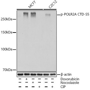
Western blot analysis of various lysates using Phospho-POLR2A CTD-S5 Rabbit mAb (CABP0997) at 1:1000 dilution. MCF7 cells were treated with Doxorubicin (0. 5 uM) at 37℃ for 24 hours or treated with nocodazole (50 ng/mL) at 37℃ for 20 hours or treated with CIP(20uL/400ul) at 37℃ for 1 hour. C2C12 cells were treated with CIP(20uL/400ul) at 37℃ for 1 hour. Secondary antibody: HRP-conjugated Goat anti-Rabbit IgG (H+L) (CABS014) at 1:10000 dilution. Lysates/proteins: 25μg per lane. Blocking buffer: 3% BSA. Detection: ECL Basic Kit (AbGn00020). Exposure time: 1s.
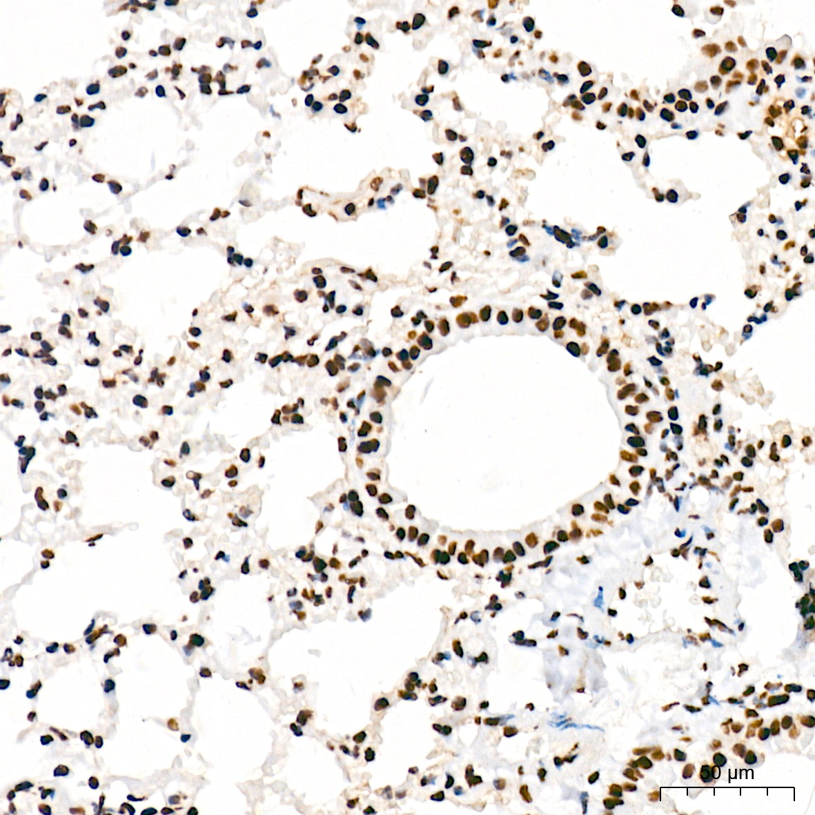
Immunohistochemistry analysis of paraffin-embedded Mouse lung tissue using Phospho-POLR2A CTD-S5 Rabbit mAb (CABP0997) at a dilution of 1:200 (40x lens). High pressure antigen retrieval was performed with 0.01 M citrate buffer (pH 6.0) prior to IHC staining.
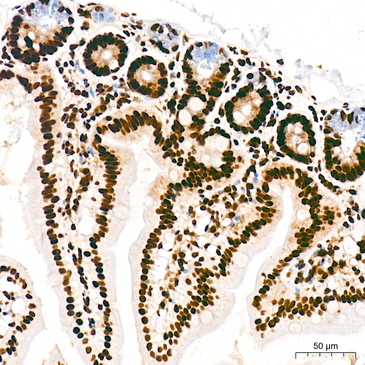
Immunohistochemistry analysis of paraffin-embedded Mouse colon tissue using Phospho-POLR2A CTD-S5 Rabbit mAb (CABP0997) at a dilution of 1:200 (40x lens). High pressure antigen retrieval was performed with 0.01 M citrate buffer (pH 6.0) prior to IHC staining.
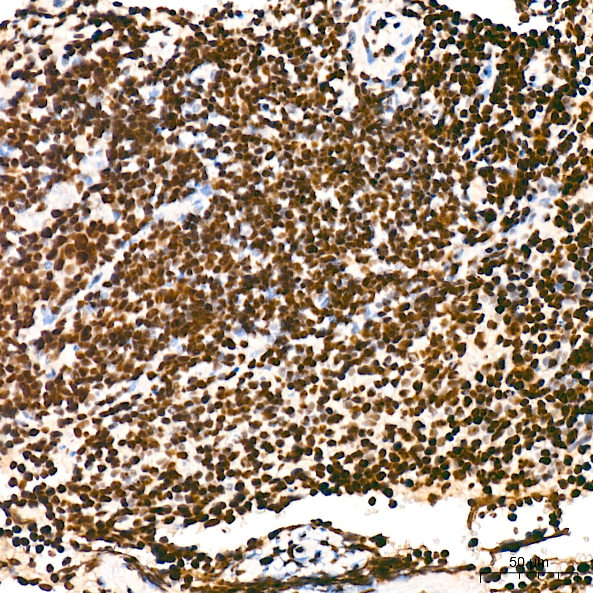
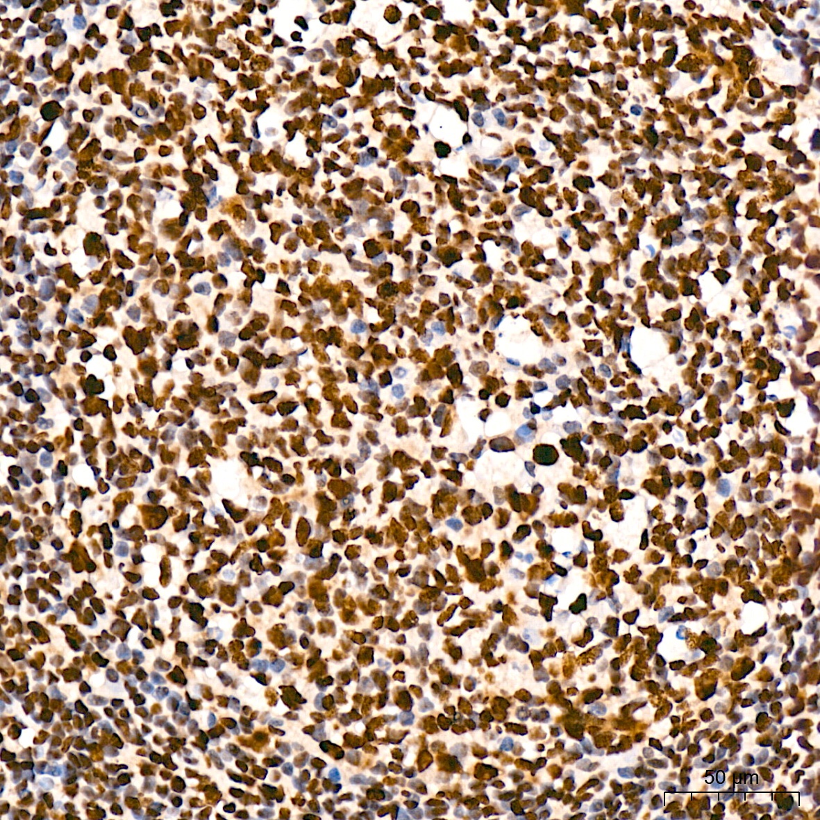
Immunohistochemistry analysis of paraffin-embedded Mouse spleen tissue using Phospho-POLR2A CTD-S5 Rabbit mAb (CABP0997) at a dilution of 1:200 (40x lens). High pressure antigen retrieval was performed with 0.01 M citrate buffer (pH 6.0) prior to IHC staining.
Immunohistochemistry analysis of paraffin-embedded Human tonsil tissue using Phospho-POLR2A CTD-S5 Rabbit mAb (CABP0997) at a dilution of 1:200 (40x lens). High pressure antigen retrieval was performed with 0.01 M citrate buffer (pH 6.0) prior to IHC staining.
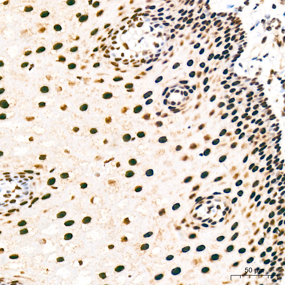
Immunohistochemistry analysis of paraffin-embedded Human esophagus tissue using Phospho-POLR2A CTD-S5 Rabbit mAb (CABP0997) at a dilution of 1:200 (40x lens). High pressure antigen retrieval was performed with 0.01 M citrate buffer (pH 6.0) prior to IHC staining.
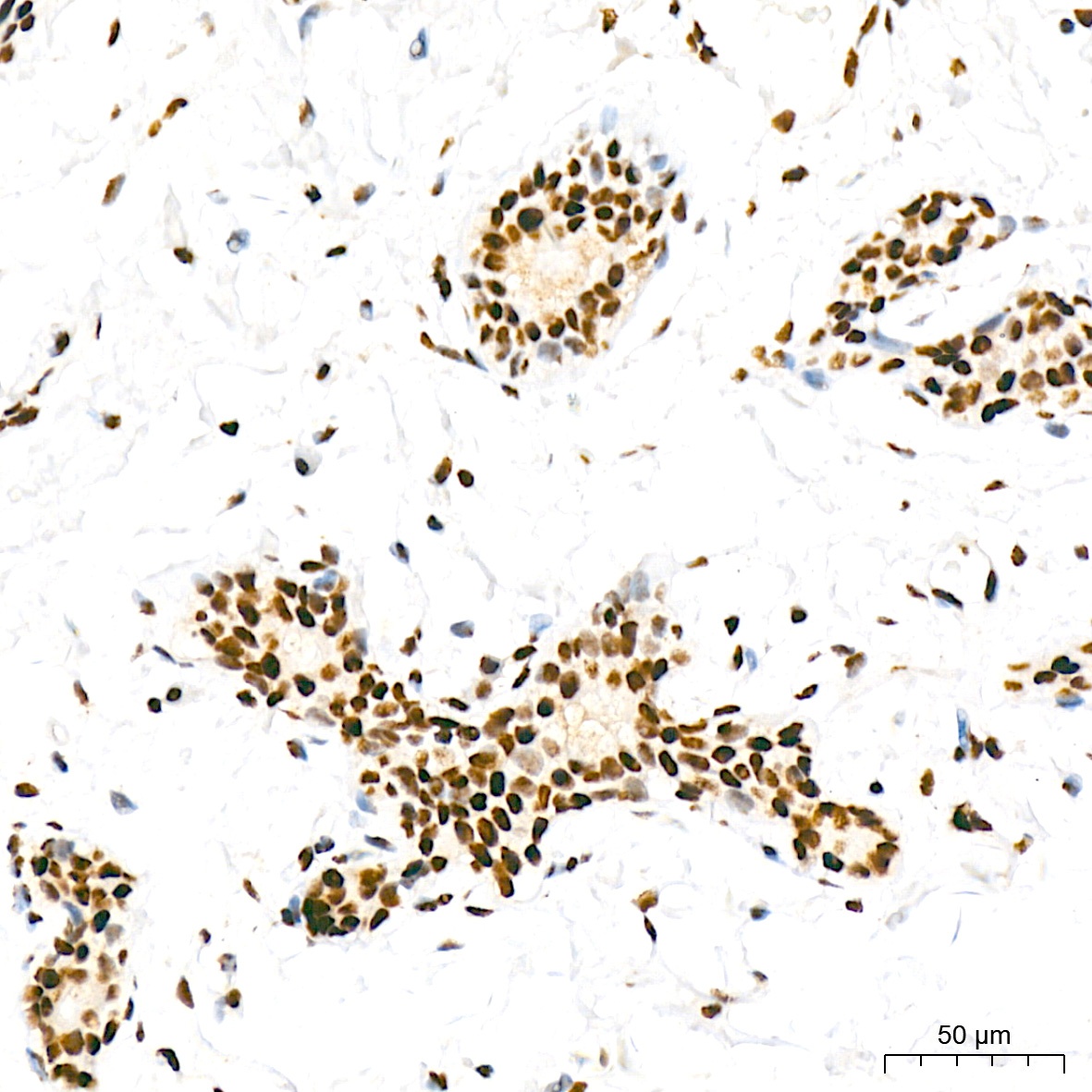
Immunohistochemistry analysis of paraffin-embedded Human breast tissue using Phospho-POLR2A CTD-S5 Rabbit mAb (CABP0997) at a dilution of 1:200 (40x lens). High pressure antigen retrieval was performed with 0.01 M citrate buffer (pH 6.0) prior to IHC staining.
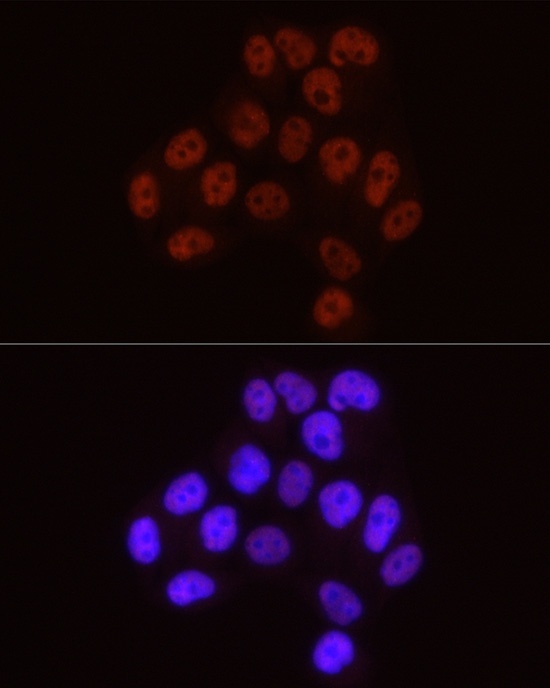
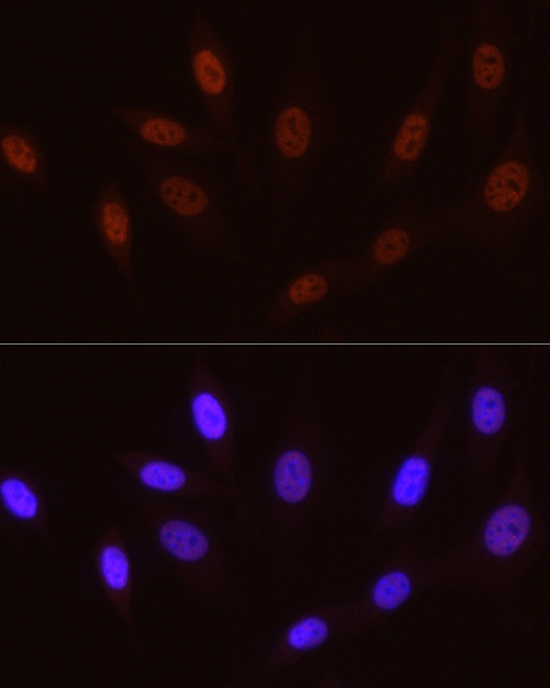
Immunofluorescence analysis of A-549 cells using Phospho-POLR2A CTD-S5 Rabbit mAb (CABP0997) at dilution of 1:50 (40x lens). Secondary antibody: Cy3-conjugated Goat anti-Rabbit IgG (H+L) (CABS007) at 1:500 dilution. Blue: DAPI for nuclear staining.
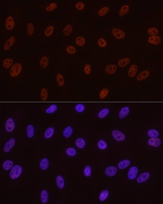
Immunofluorescence analysis of HeLa cells using Phospho-POLR2A CTD-S5 Rabbit mAb (CABP0997) at dilution of 1:50 (40x lens). Secondary antibody: Cy3-conjugated Goat anti-Rabbit IgG (H+L) (CABS007) at 1:500 dilution. Blue: DAPI for nuclear staining.
Immunofluorescence analysis of NIH/3T3 cells using Phospho-POLR2A CTD-S5 Rabbit mAb (CABP0997) at dilution of 1:50 (40x lens). Secondary antibody: Cy3-conjugated Goat anti-Rabbit IgG (H+L) (CABS007) at 1:500 dilution. Blue: DAPI for nuclear staining.


![Anti-Phospho-POLR2A (Ser5) [R08-4O2] Monoclonal Antibody (AGMB05320)](https://cdn11.bigcommerce.com/s-h68l9z2lnx/images/stencil/590x590/products/276605/676856/anti-phospho-polr2a-ser5-r08-4o2-monoclonal-antibody-agmb05320__76671.1773030749.jpg?c=2 "Anti-Phospho-POLR2A (Ser5) [R08-4O2] Monoclonal Antibody (AGMB05320)")